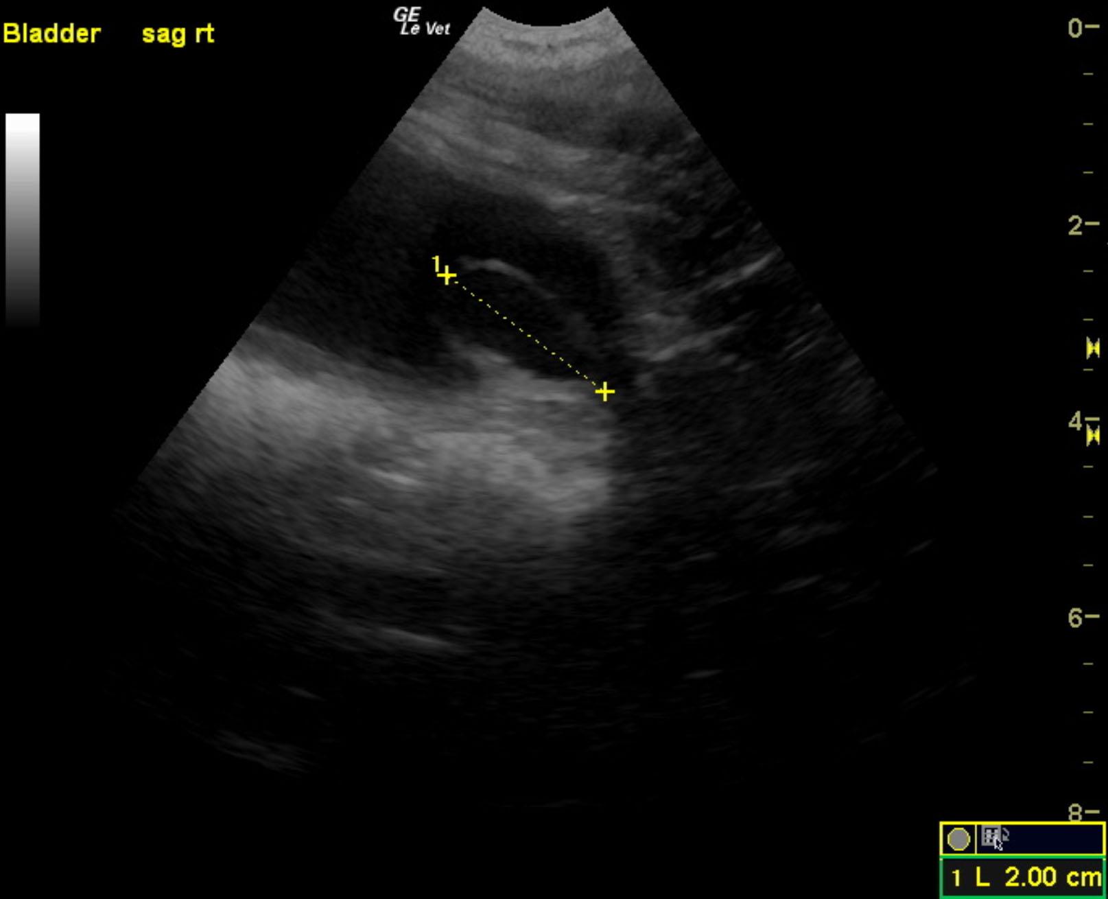
A 3-year-old neutered male Labrador Retriever dog was presented for evaluation of decreased appetite, possible polyuria and polydypsia, taking longer to urinate than normal, and hematuria. On urinalysis, isosthenuria (1.016), 3+ protein, white blood cells, and red blood cells were present. Stress leukogram was evident on CBC, but serum chemistry was within normal limits.
A 3-year-old neutered male Labrador Retriever dog was presented for evaluation of decreased appetite, possible polyuria and polydypsia, taking longer to urinate than normal, and hematuria. On urinalysis, isosthenuria (1.016), 3+ protein, white blood cells, and red blood cells were present. Stress leukogram was evident on CBC, but serum chemistry was within normal limits.