06_00135 Patches S Suspect interstitial cystitis

Case Study

06_00135 Patches S Suspect interstitial cystitis

A 4-year-old SF DSH was presented for evaluation of  dysuria, stranguria, and hematuria over a 5-day period that had not responded to antibiotics. Urinalysis revealed pH of 8, 2+ protein and 3+ blood. 
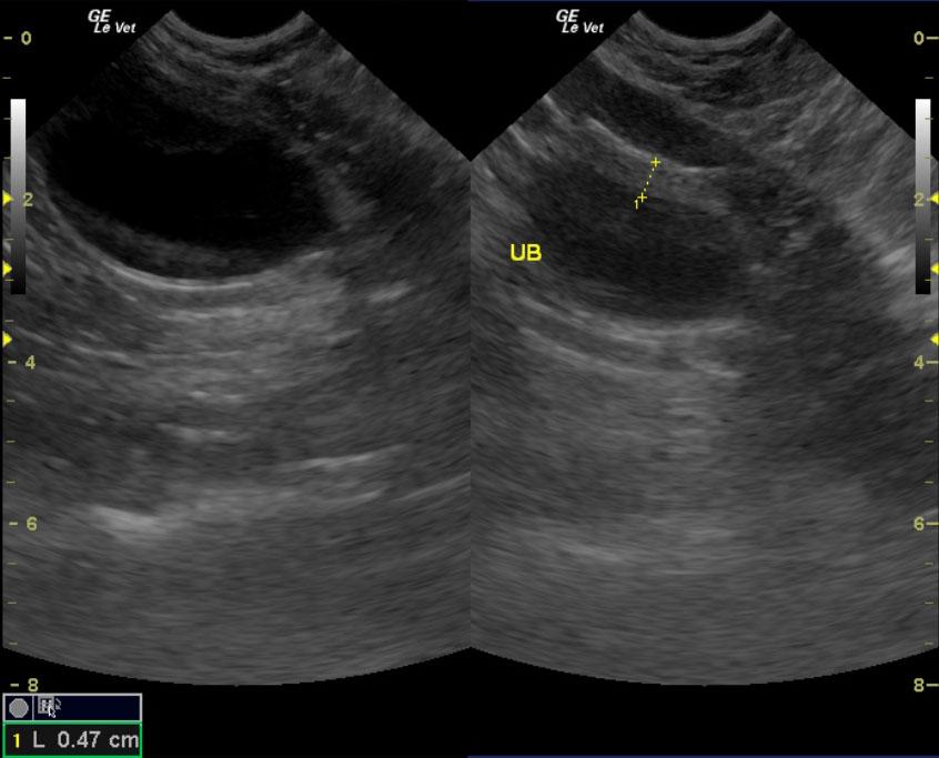
A 4-year-old SF DSH was presented for evaluation of  dysuria, stranguria, and hematuria over a 5-day period that had not responded to antibiotics. Urinalysis revealed pH of 8, 2+ protein and 3+ blood. 
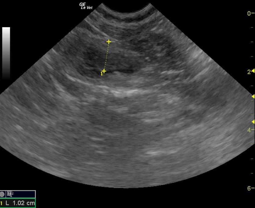
Sonographic Differential Diagnosis

Focally thickened bladder wall with loss of detail in the urinary bladder with echogenic polypoid changes. I strongly recommend surgical biopsies or debulking of the bladder wall in this patient with appropriate biopsies and culture. Severe interstitial cystitis is suspected with the possibility of bladder lymphoma. Traumatic catheterization is also a possibility. FNA with cytology can also be considered. Guarded prognosis.

Image Interpretation

The urinary bladder was focally thickened and empty at the time of the sonogram and measured 0.93 cm. Focal, irregular thickening of the ventral wall was noted. No evidence of calculi was present at the time of the sonogram. Significant loss of detail was noted in the bladder wall itself with echogenic, polypoid changes. The dorsal wall appeared more regular, yet micropolypoid changes were present.

DX

Suspect interstitial cystitis

Outcome

The patient is currently doing well and urinating normally.

Clinical Differential Diagnosis

Interstitial cystitis, uroliths, neoplasia, bacterial cystitis, granulomatous cystitis

UA PH

8

UA PH

8

Sampling

FNA of the bladder mucosa revealed poor exfoliation.

Patient Information

Gender : Female, Spayed
Species : Feline
Type of Imaging : Ultrasound

Clinical Signs

  • Dysuria
  • Hematuria
  • Stranguria

Images

wall_measempty_meas

Clinical Signs

  • Dysuria
  • Hematuria
  • Stranguria

Urinalysi

  • Blood Present
  • pH High
  • Protein Present