06_00124 Dakota G Renal mass

Case Study

06_00124 Dakota G Renal mass

A 10-year-old SF DSH was presented for evaluation of ravenous appetite and weight loss. On physical examination an enlarged right kidney and thickened bowel loops were present. Leukocytosis was evident at 26,000.
A 10-year-old SF DSH was presented for evaluation of ravenous appetite and weight loss. On physical examination an enlarged right kidney and thickened bowel loops were present. Leukocytosis was evident at 26,000.
Sonographic Differential Diagnosis

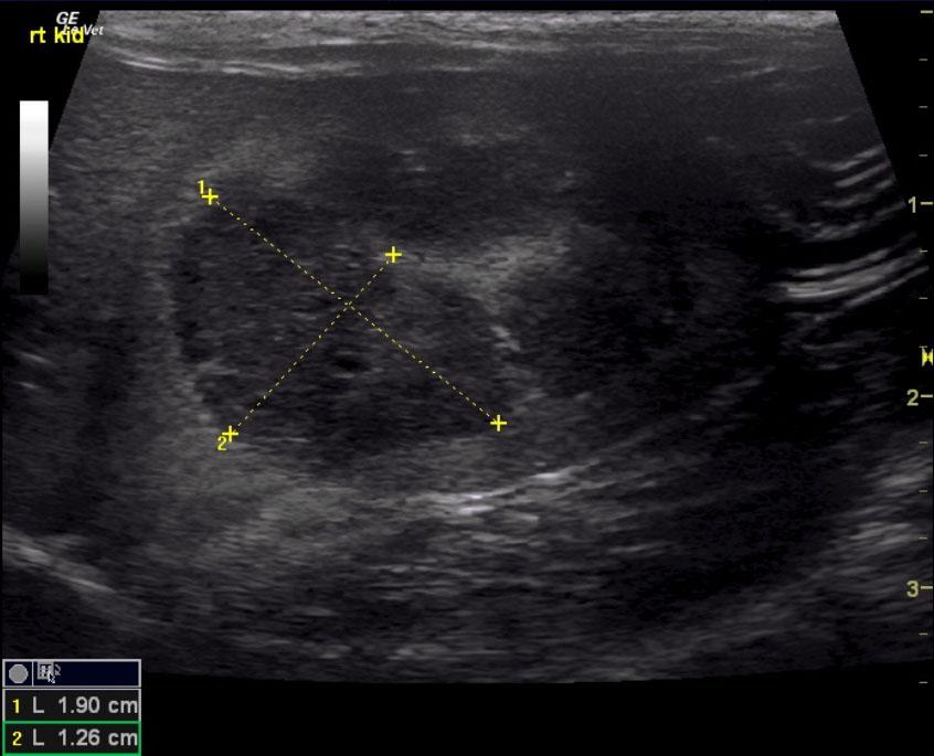
Right kidney mass. May represent necrotic abscessation given the calculus. Passage of calculus may predispose to an abscess or more likely represent a neoplastic event.
The left kidney appears structurally viable enough to sustain metabolic need.
Ultrasound-guided FNA +/- culture and cytology would be recommended or right nephrectomy.

Image Interpretation

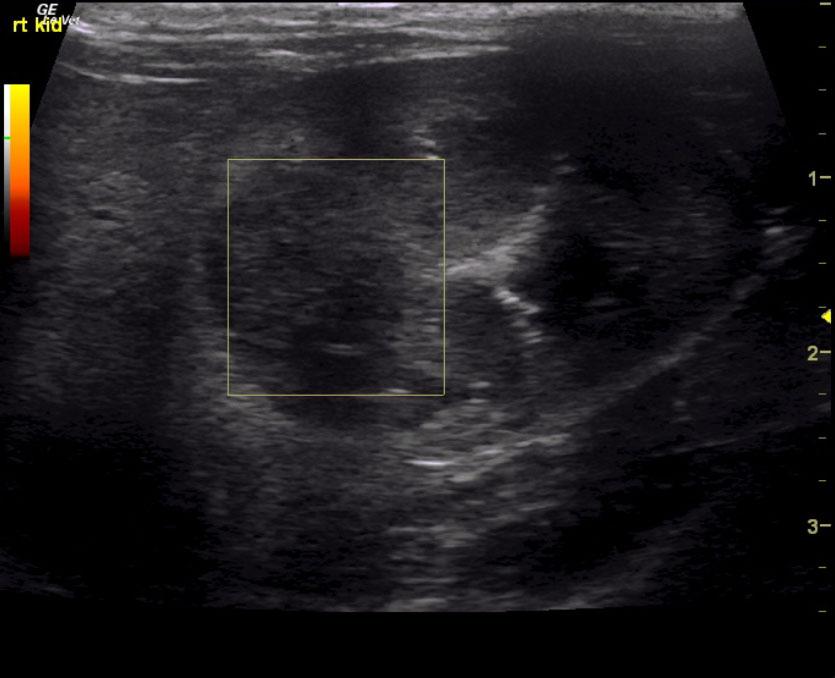
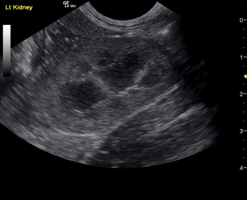
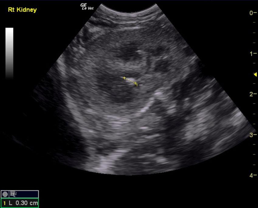
The left kidney was uniform, without calculus, and measured 3.75 cm. The cranial pole of the right kidney presented a hypoechoic mass that measured 1.9 x 1.26 cm with disruption of the corticomedullary junction and capsular swelling. There was no blood flow to the mass. The right kidney presented a calculus at the corticomedullary junction. The right kidney measured 3.76 cm. FNA of the right kidney is recommended.

DX

Renal mass

Outcome

None

Clinical Differential Diagnosis

Hyperthyroidism
Renal – neoplasia, cyst, hydronephrosis, renolith, abscess
Bowel – IBD, neoplasia, granulomatous disease, foreign body

Sampling

None

Patient Information

Gender : Female, Spayed
Species : Feline
Type of Imaging : Ultrasound
Status : Complete

Clinical Signs

  • Weight loss

Exam Finding

  • Large Kidneys
  • Thickened Intestines

Images

lkrtcalcmeaspwr_dop

CBC

  • WBC, High

Clinical Signs

  • Weight loss