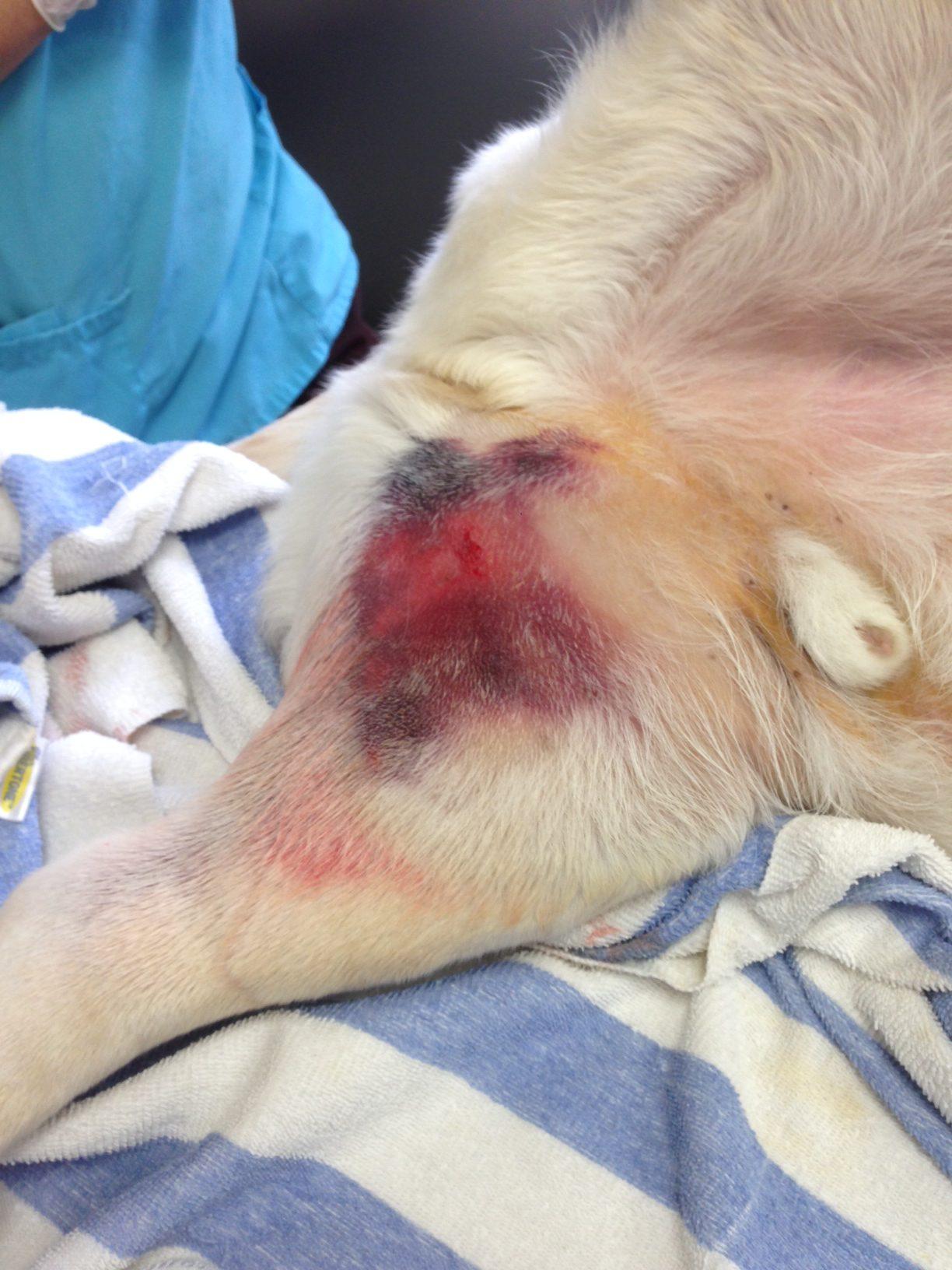
A 7-year-old MN Labrador Retriever was presented for severe hind limb swelling, bruising in the inguinal region (Image 1) and grade 3 limp. Mild fever was present. Radiographs revealed severe soft tissue swelling of around the femur without bone involvement. Thoracic and abdominal radiographs were unremarkable.
A 7-year-old MN Labrador Retriever was presented for severe hind limb swelling, bruising in the inguinal region (Image 1) and grade 3 limp. Mild fever was present. Radiographs revealed severe soft tissue swelling of around the femur without bone involvement. Thoracic and abdominal radiographs were unremarkable.