Sarcoma of the medial aspect of the forelimb and regional lymphadenopathy in a 7 year old MN Corgi dog.

Case Study

Sarcoma of the medial aspect of the forelimb and regional lymphadenopathy in a 7 year old MN Corgi dog.

A 7-year-old male neutered Corgi with a history of lameness and osteoarthritis was presented for evaluation of a cavitated mass on the front leg with prescapular lymphadenomegaly.
A 7-year-old male neutered Corgi with a history of lameness and osteoarthritis was presented for evaluation of a cavitated mass on the front leg with prescapular lymphadenomegaly.
Sonographic Differential Diagnosis

Soft tissue mass in the right leg with regional lymphadenopathy. This presentation is strongly suggestive for sarcoma or similar neoplasia.

Image Interpretation

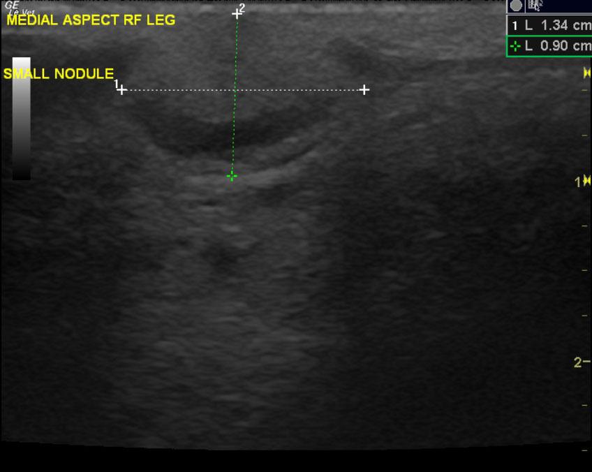
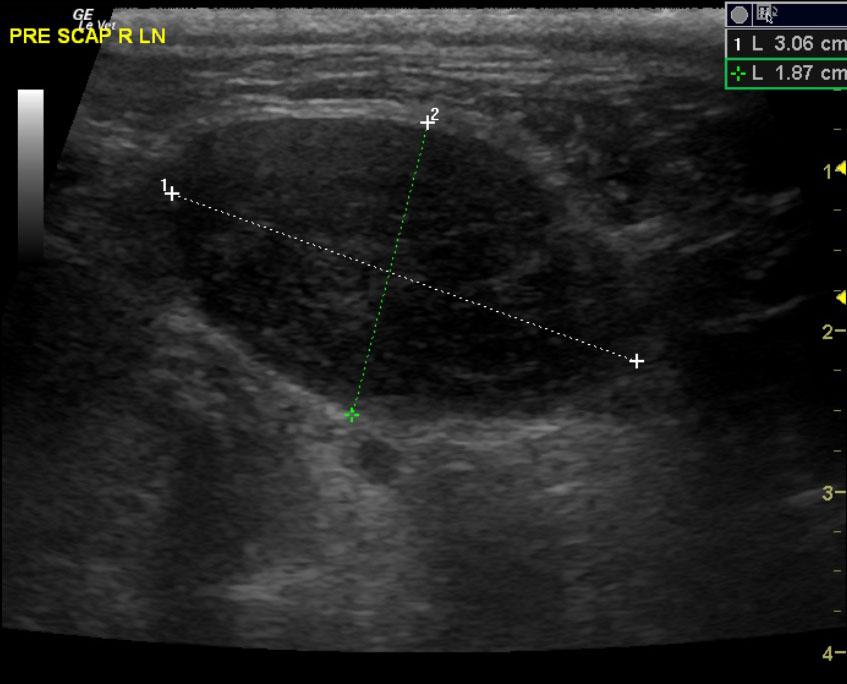
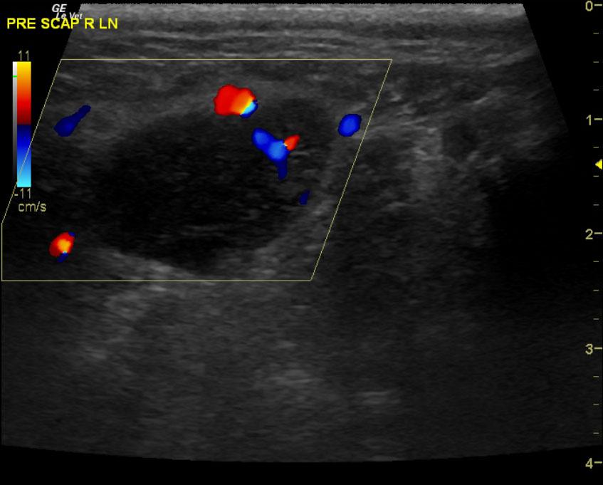
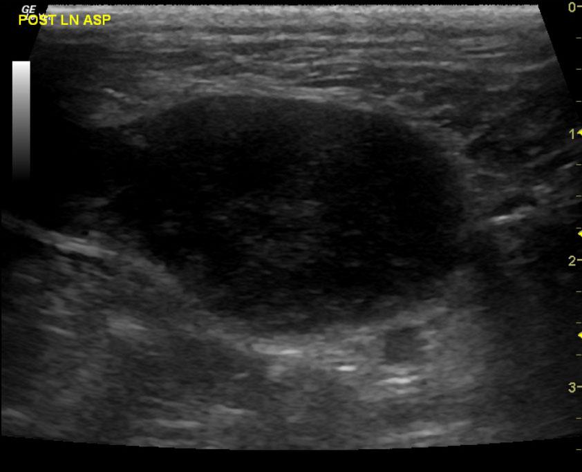
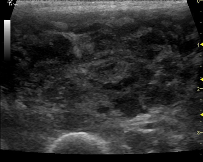
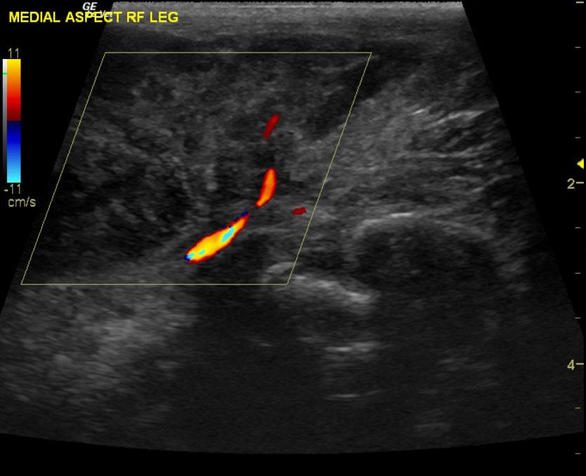
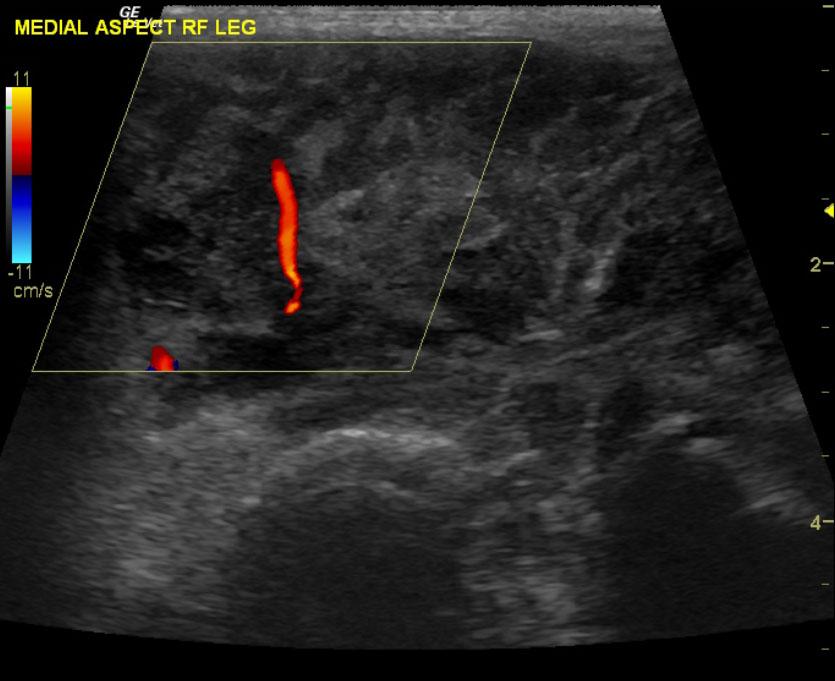
The right forelimb in this patient presented a mixed, hypoechoic, micronodular mass with dramatic hypoechoic regions. This mass appeared to be of soft tissue origin in the right leg and appears to be significantly vascular on color flow Doppler. The bone does not appear to be involved as the periosteum appears normal. The regional prescapular lymph node was enlarged, hypoechoic and irregular. This is strongly suggestive for infiltrative disease and measured 3.06 x 1.87 cm. A second soft tissue nodule was noted on the medial aspect of the right leg as well with a similar regional inflammatory pattern.

DX

Leg mass: sarcoma

Outcome

None

Comments

Abdominal and thoracic ultrasound is recommended for staging purposes. Oncological consultation is recommended; amputation of the leg is likely outcome. However, given the lymph node spread, multicentric infiltrative disease may be present. Very guarded to poor prognosis.

Clinical Differential Diagnosis

Neoplasia, abscess, granuloma, cyst

Sampling

FNAs were performed of the lymph node and the mass itself without complication. Cytology revealed sarcoma and possibilities included soft tissue sarcoma or histiocytic sarcoma. Cytology of the lymph node revealed marked suppurative inflammation with evidence of tissue necrosis.

Patient Information

Patient Name : Liam M
Gender : Male, Neutered
Species : Canine
Type of Imaging : Ultrasound
Status : Complete
Liz Wuz Here : Yes
Code : 13_00016

Clinical Signs

  • Enlarged Lymph Nodes
  • Lameness

Images

mass_1mass_d_1mass_d_2mass_2lnln_dln_post

Clinical Signs

  • Enlarged Lymph Nodes
  • Lameness