A 10-year-old MN DSH cat was presented for evaluation of lethargy and poor appetite for the past week. Weight loss was evident and although there was no mention of vomiting, the patient did vomit whilst hospitalized overnight. Abnormalities on CBC and serum biochemistry were neutrophilia, lymphopenia, and mild hyponatremia. Dilated stomach and a full and possibly plicated colon were evident on survey radiographs.
A 10-year-old MN DSH cat was presented for evaluation of lethargy and poor appetite for the past week. Weight loss was evident and although there was no mention of vomiting, the patient did vomit whilst hospitalized overnight. Abnormalities on CBC and serum biochemistry were neutrophilia, lymphopenia, and mild hyponatremia. Dilated stomach and a full and possibly plicated colon were evident on survey radiographs.
Case Study
Obstructive intestinal masses in a 10-year-old MN DSH cat
Sonographic Differential Diagnosis
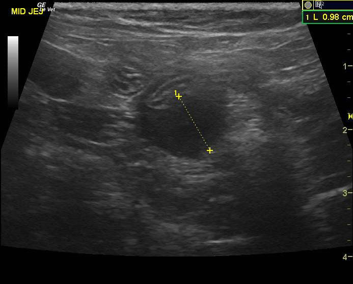
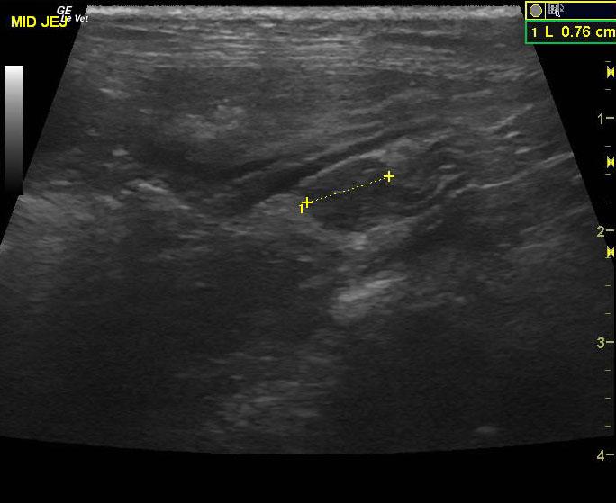
Distal small intestinal foreign body obstruction with concurrent focal, intestinal mass that measured 1.5 x 2.0 cm. The intestinal lesion is strongly suggestive of lymphoma, possible mast cell disease, FIP or less likely carcinoma.
Mesenteric lymph node pathology – lymphadenitis, neoplasia
Image Interpretation
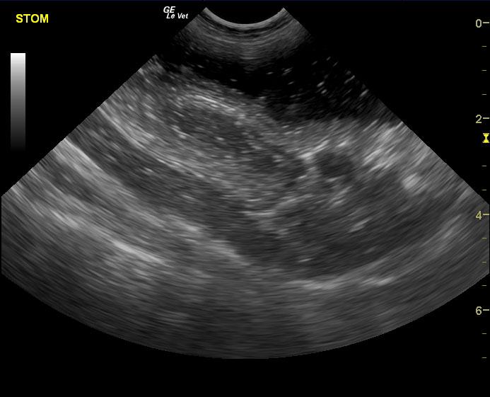
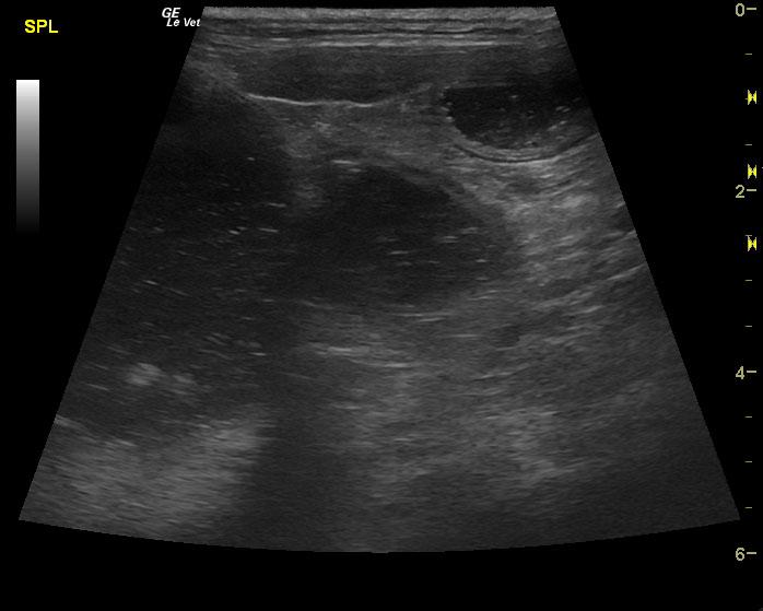
The stomach in this patient was severely dilated as was the small intestine. A hardened and progressively shadowing 2.0 cm foreign body was noted in the distal small intestine and caused an obstructive pattern. Some aspects of a potential linear accumulation were noted. A separate area of jejunum presented a hypoechoic, irregular wall deriving from the muscularis with loss of structural detail. . The mesenteric root lymph nodes were mildly prominent and uniform. Focal, reactive surrounding omentum was also noted.
DX
Outcome
Owners elected exploratory. There were 3 approximately 1 cm masses in the distal jejunum about 4 cm apart. One or more appeared to be large enough to occlude the lumen. There was a large bundle of enlarged lymph nodes in the mesentery area. The entire area was resected, distal section turned muddy red – discussed intraoperative euthanasia with owner. Elected to wake the patient up and see how it goes. Cat is doing better. Elected to wait on sending in biopsy.
Comments
Foreign body removal, intestinal resection, which is a separate portion of the obstruction would be recommended with variable intestinal wall biopsies and biopsies of masses. Intraoperative ultrasound would be ideal in this case. Biopsy of the reactive mesenteric lymph node would be recommended at the time of surgery
Clinical Differential Diagnosis
GI tract pathology – obstruction (neoplasia/foreign body), IBD, neoplasia, constipation, granulomatous disease; Pancreatic pathology – pancreatitis, neoplasia; FIP, Neoplasia
Sampling
biopsy samples obtained of the masses and mesenteric lymph nodes; o elected not to send to lab.
Video
Patient Information
Clinical Signs
- Anorexia
- Lethargy
Exam Finding
- Vomiting
- Weight loss
Images
Blood Chemistry
- Sodium, Low
CBC
- Lymphocytes, Low
- Neutrophils, High
Clinical Signs
- Anorexia
- Lethargy