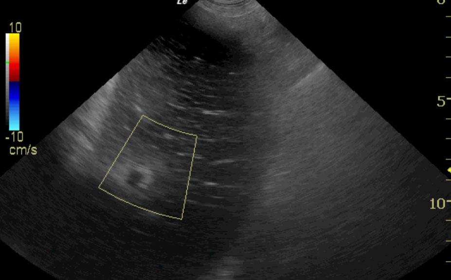
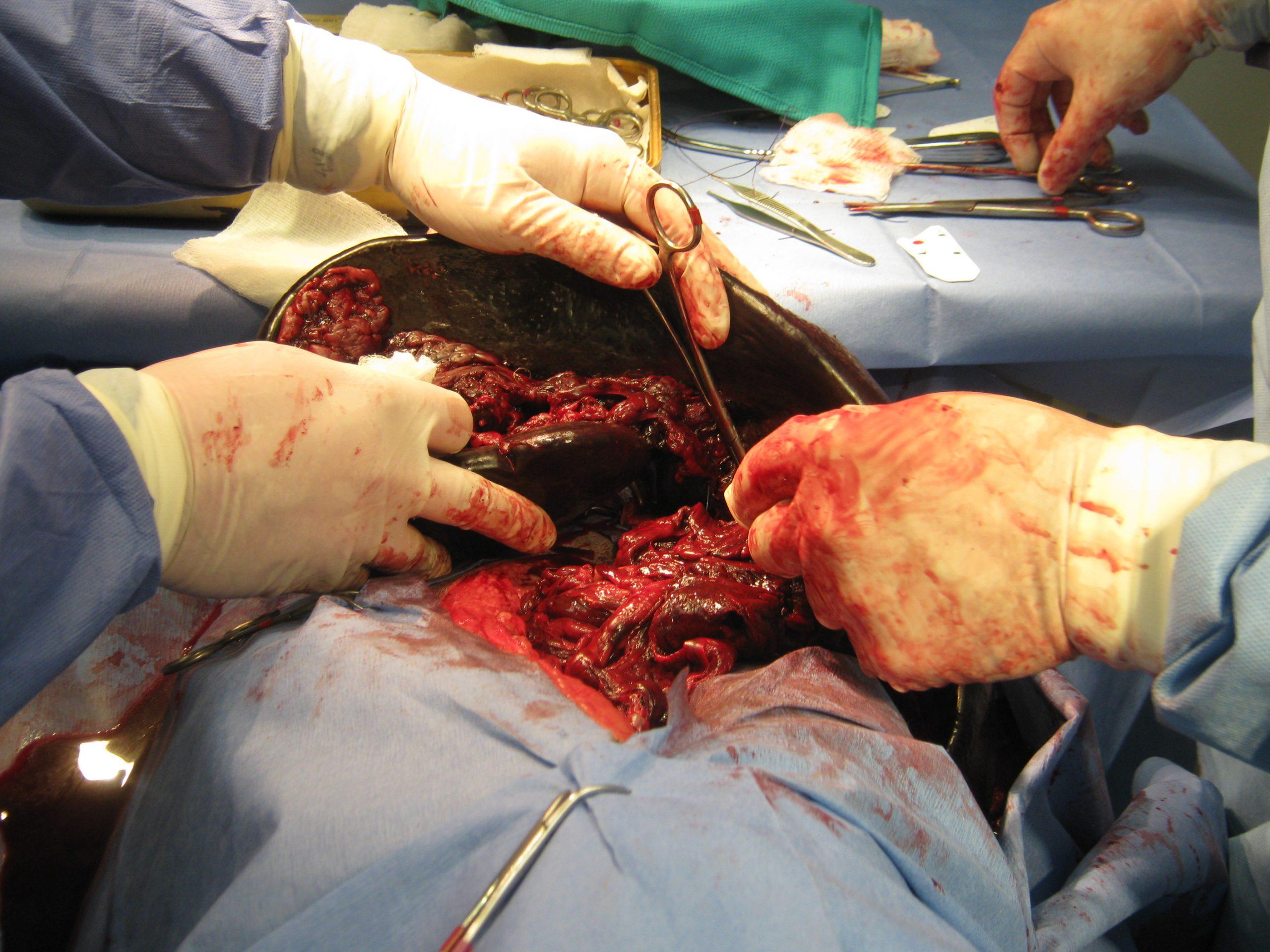
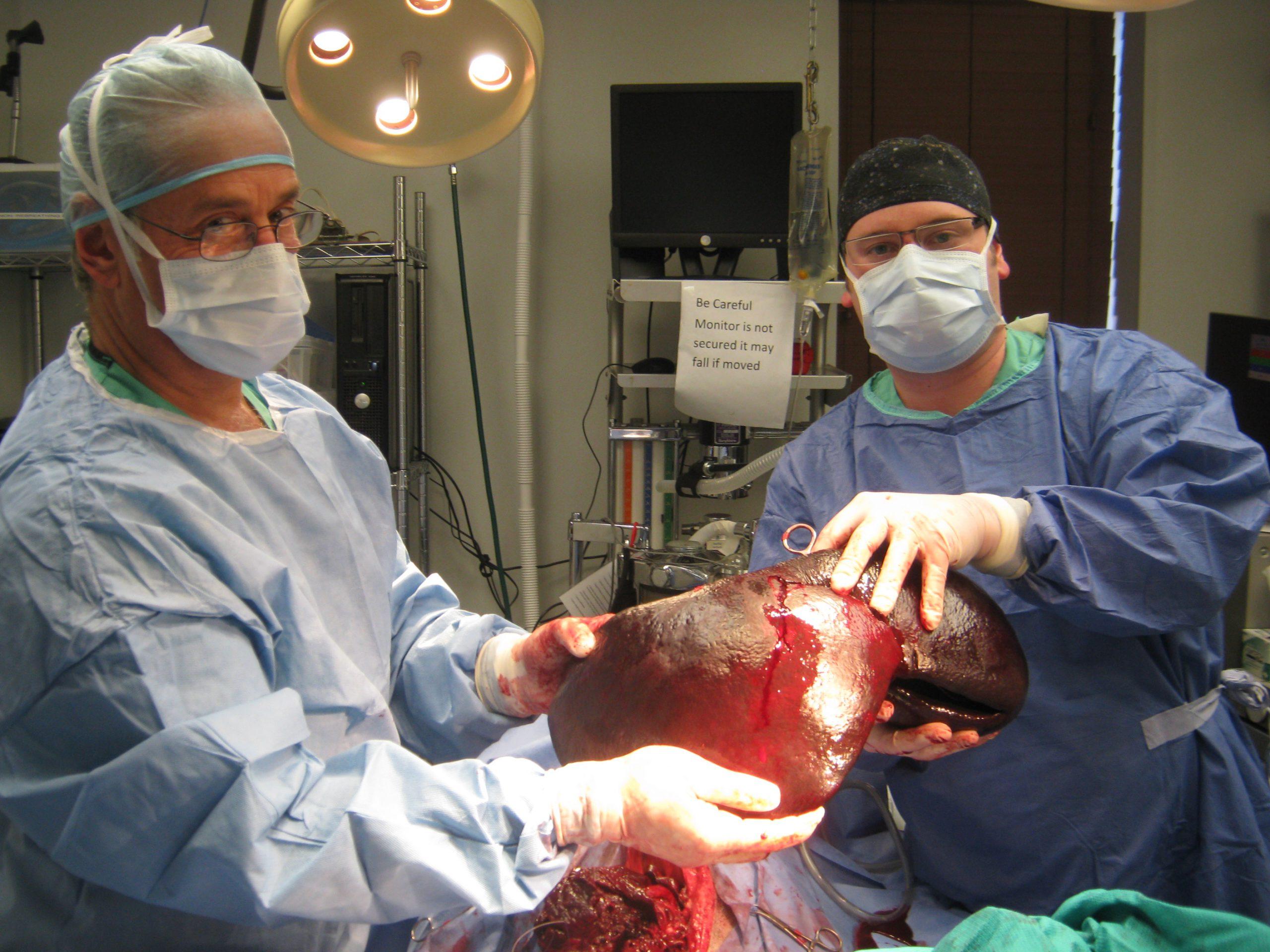
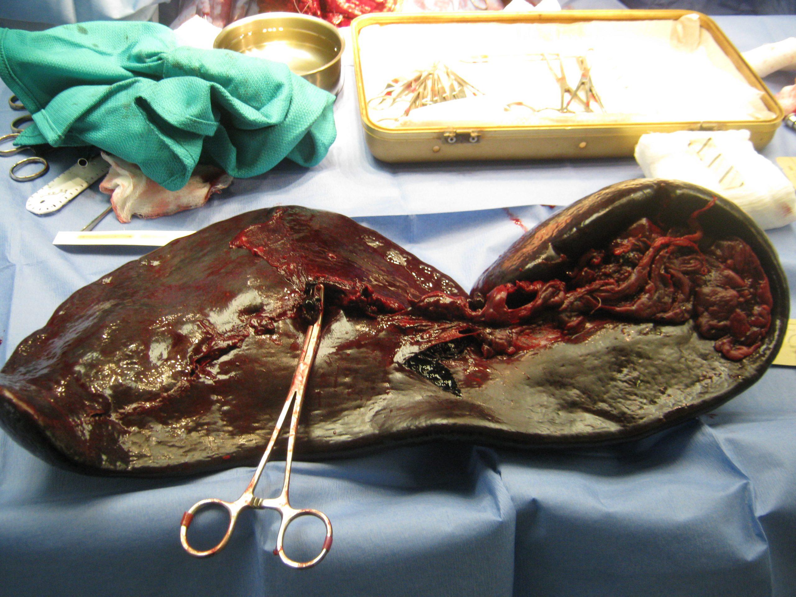
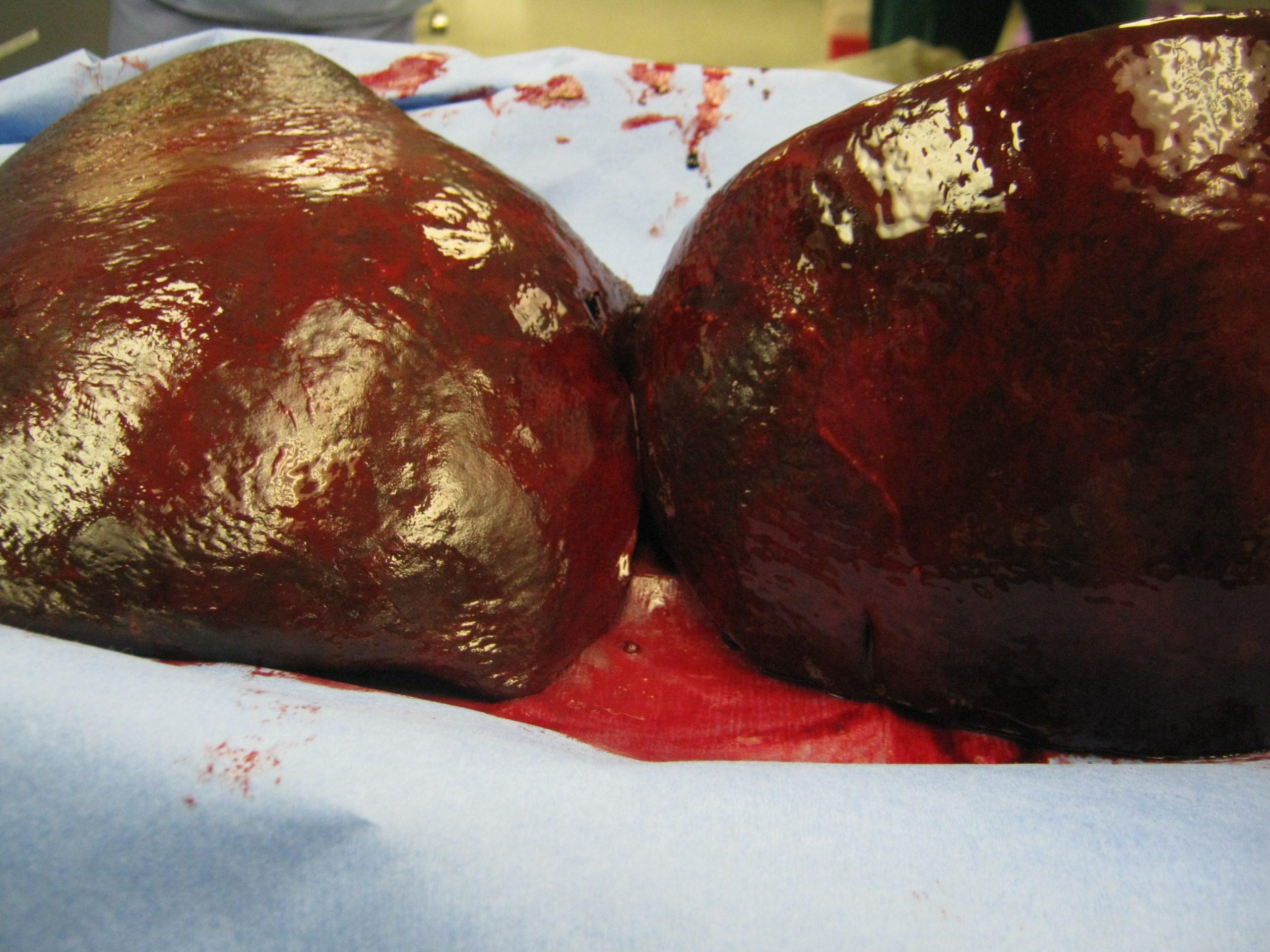
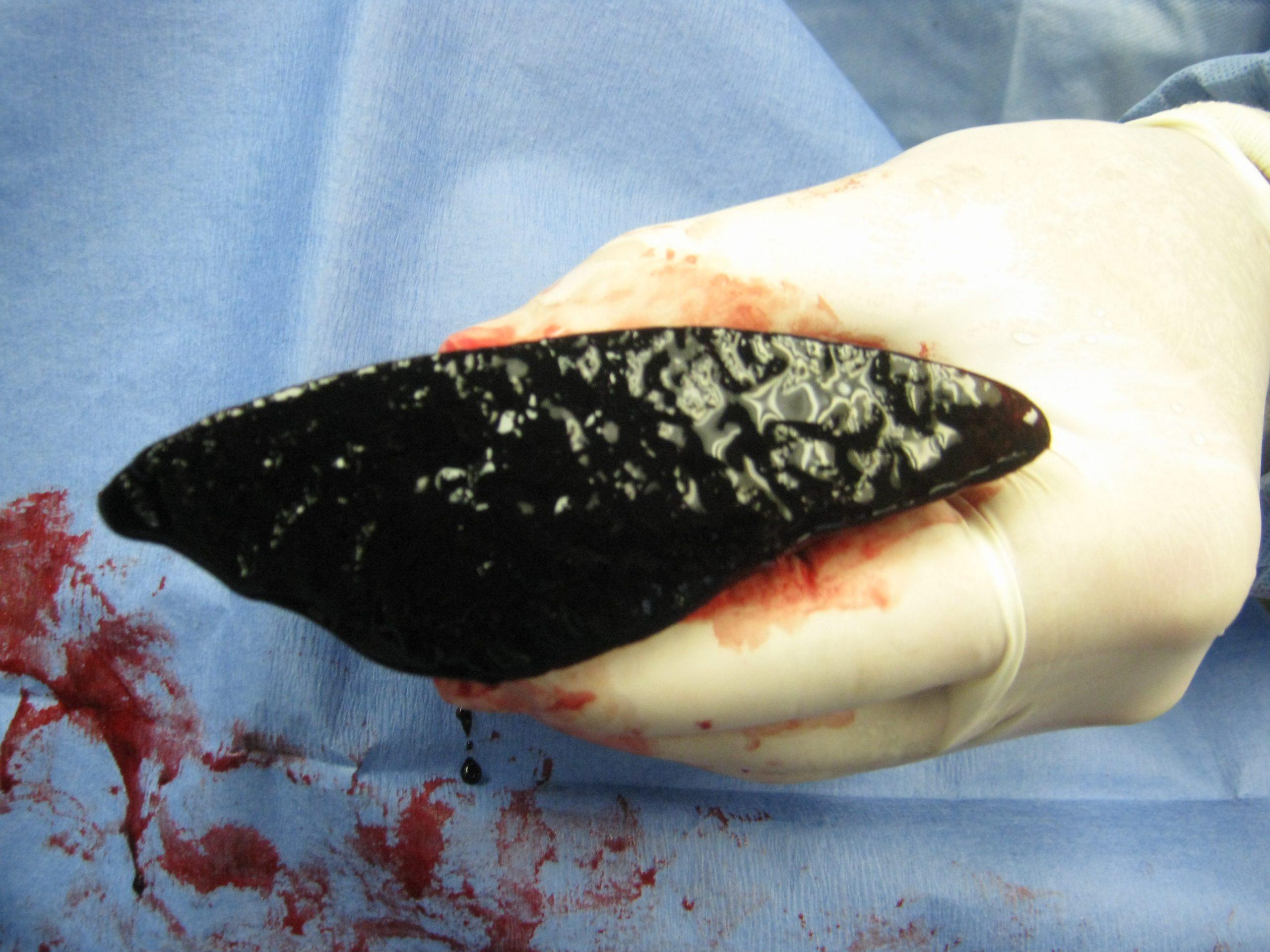
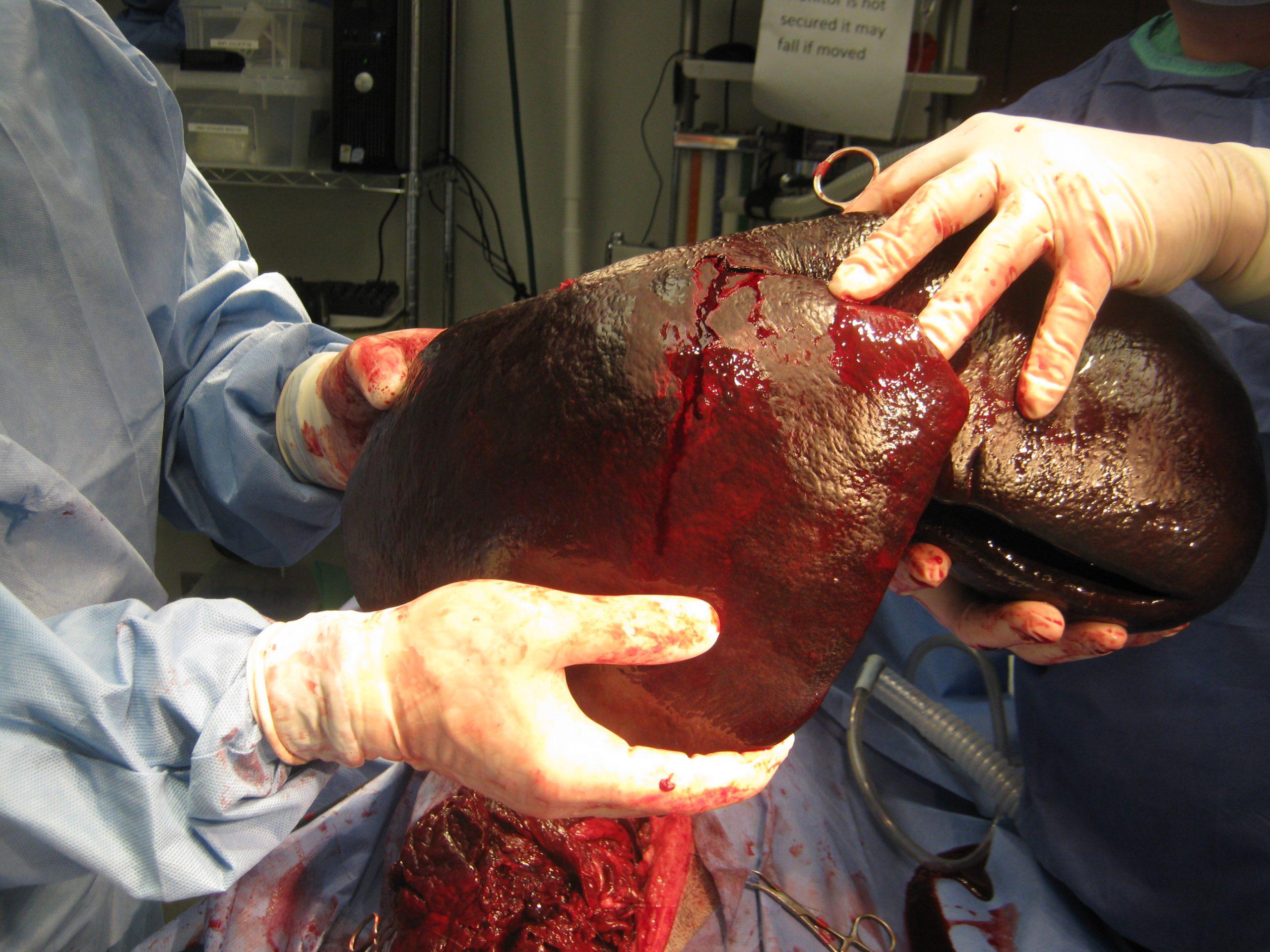
An intact 7-year-old female Great Dane dog presented for a history of vomiting and lethargy over a couple of days. At the previous assessment, anemia (32.5%), leukocytosis, and free fluid were present. On follow up assessment, the anemia had worsened (27.5%) and the leukocytosis was similar. ECG revealed VPCs. On cursory abdominal ultrasonography, free fluid and hyperechoic hepatic nodules were evident. The free fluid was found to represent a hemoabdomen.
An intact 7-year-old female Great Dane dog presented for a history of vomiting and lethargy over a couple of days. At the previous assessment, anemia (32.5%), leukocytosis, and free fluid were present. On follow up assessment, the anemia had worsened (27.5%) and the leukocytosis was similar. ECG revealed VPCs. On cursory abdominal ultrasonography, free fluid and hyperechoic hepatic nodules were evident. The free fluid was found to represent a hemoabdomen.