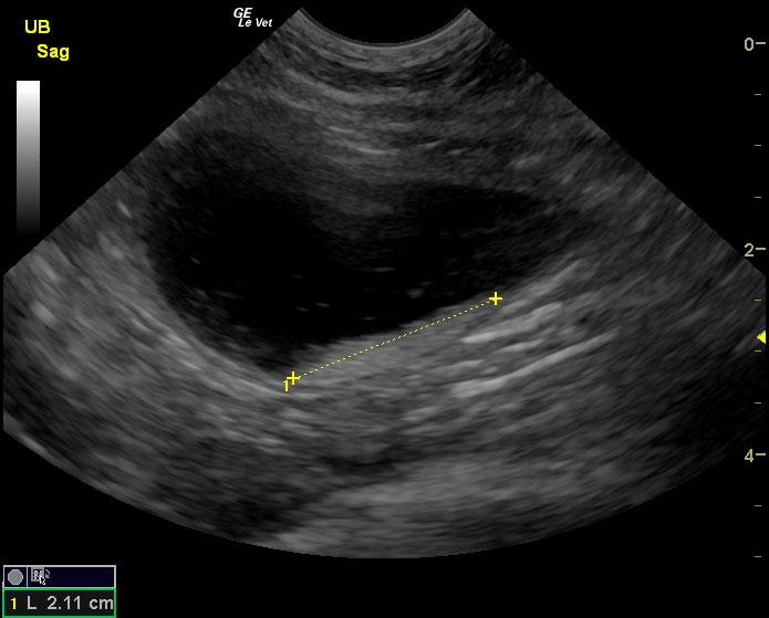
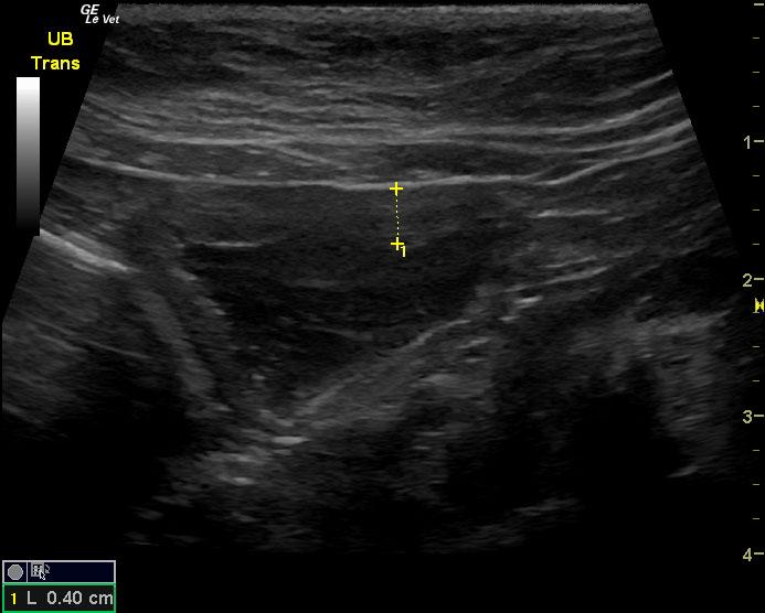
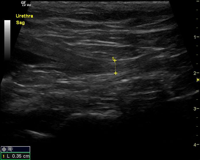
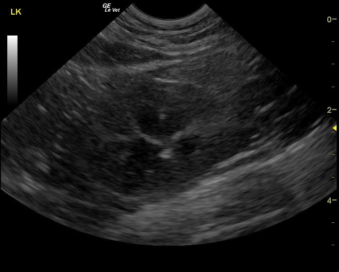
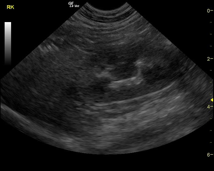
The urinary bladder in this patient revealed dependent debris that measured 2.1 cm in length. The bladder wall presented minor, apical ventral wall thickening. Suspended debris was also evident. The bladder wall thickening appeared to be concentric throughout with some loss of mural detail, and measured 0.4 cm at minor repletion. The pelvic urethra was also thickened. These images are most consistent with interstitial cystitis with minor potential for bladder lymphoma. The kidneys revealed largely normal size and structure; corticomedullary definition and ratio (cortex 1/3 of medulla) were essentially maintained with minor loss of curvilinear pattern. The cortices presented largely uniform texture with some echogenic changes that are not likely of clinical significance at this time.