Cushing`s disease in a 10 year old FS Burmese cat

Case Study

Cushing`s disease in a 10 year old FS Burmese cat

A 10-year-old FS Burmese cat was presented for evaluation of possible Cushing’s disease; the cat exhibited chronic skin disease, hyperglycemia, and glucosuria but normal fructosamine.
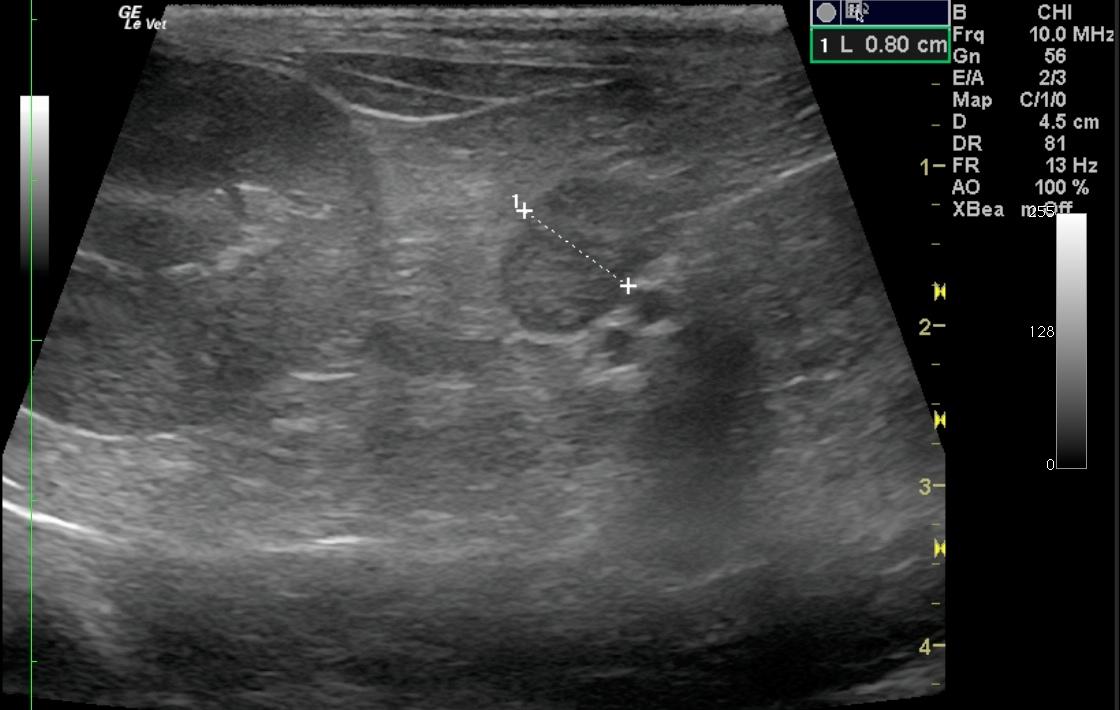
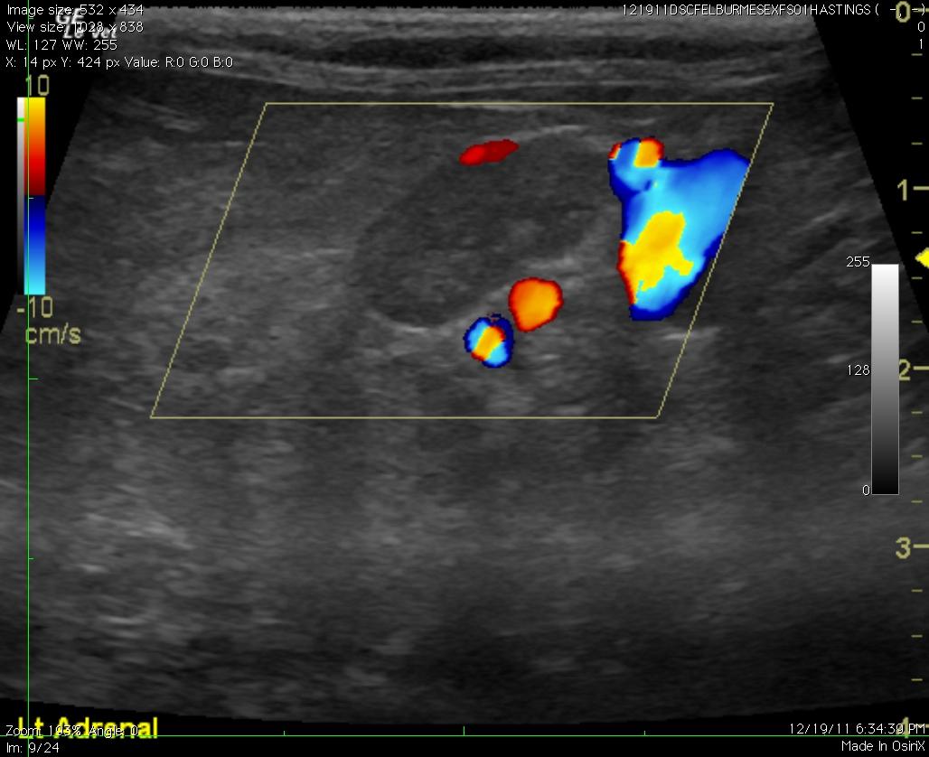
A 10-year-old FS Burmese cat was presented for evaluation of possible Cushing’s disease; the cat exhibited chronic skin disease, hyperglycemia, and glucosuria but normal fructosamine.
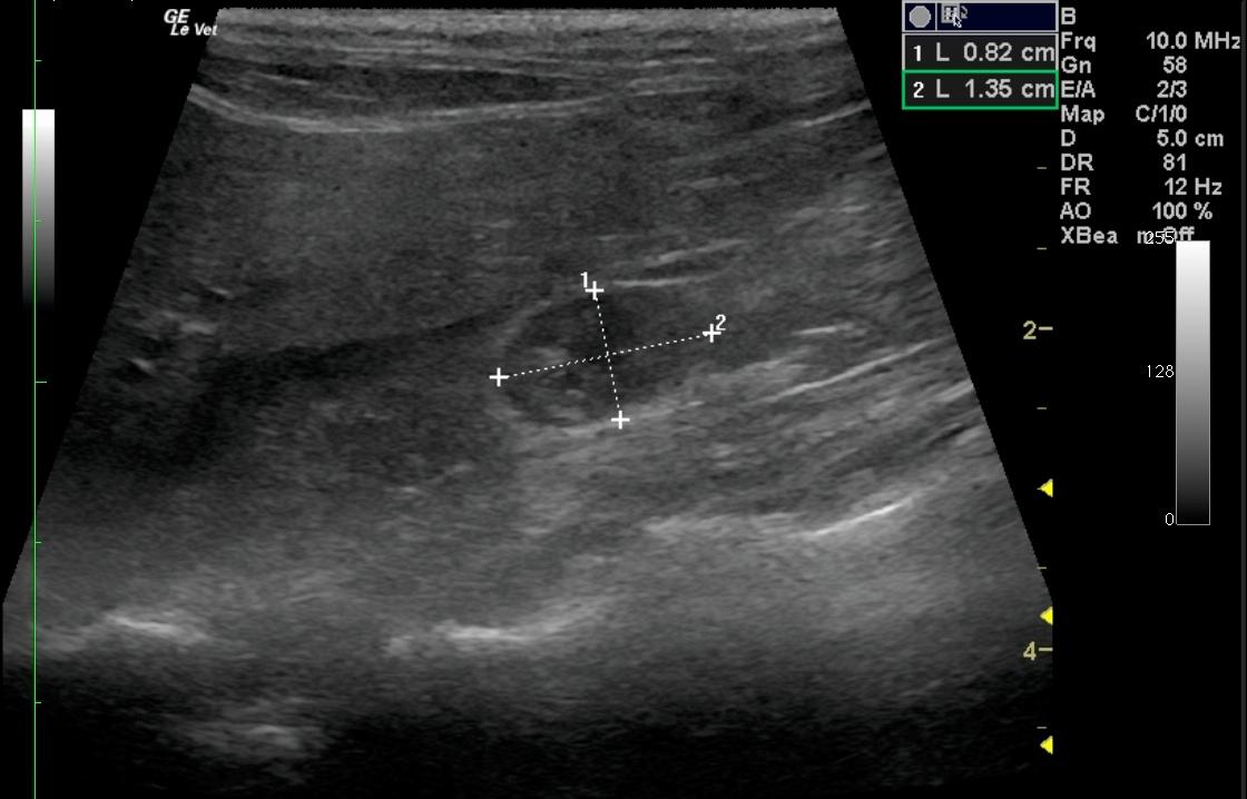
Sonographic Differential Diagnosis

Bilateral adrenal gland enlargement. Adrenal hyperplasia is suspected, particularly central Cushing’s disease and/or potential underlying acromegaly. There is a lower likelihood of stress induced hyperplasia. Concurrent low-grade chronic active pancreatitis is evident, with nodular hyperplasia and mild potential for emerging pancreatic neoplasia. The hyperechoic liver is consistent with lipidosis, biliary calculosis and potential low-grade inflammatory hepatopathy.

Image Interpretation

The left adrenal gland was moderately enlarged at 1.4 x 0.9 cm. The right adrenal gland was also uniformly enlarged and measured 1.35 x 0.82 cm. Minor parenchyma heterogenicity was noted in both glands without mass effects on the adrenal capsules. The liver is uniformly hyperechoic with normal gall bladder contour and distension (Video 3). Focal areas of lobar mineralization were noted consistent with biliary calculi. The pancreas presented heterogenic mixed hypoechoic nodular changes with minor capsular expansion (Video 4). Echogenic remodeling is also noted with undulating capsular contour.

DX

Cushing`s disease

Outcome

Trilostane and primary diabetic management were recommended. Owners elected humane euthanasia.

Comments

Central Cushing’s disease in a cat. No trouble finding the adrenals on this feline as our SonoPath collaborator and fellow Dip. ABVP, Dr. Doug Casey of English Bay Ultrasound Services in Vancouver, BC, Canada (http://www.sonopath.com/specialists_casey.asp), shows us in this SonoPath case of the month. Now we bring you a cat with central Cushing’s and, of course, a little lipidosis and pancreatitis to round out the clinical profile in this patient. In October 2011 we brought you a Cushing’s cat with an invasive adrenal mass that you may compare this case to from our past cases of the month line-up (http://www.sonopath.com/resources_archived_cases.asp). Also check out Doug’s folder of utility medicine full of things that are easy and for practical use in small animal medicine that is now live with the rest of our specialty folders.

Clinical Differential Diagnosis

Early diabetes mellitus, acromegaly, hyperthyroidism, Cushing’s disease, drugs, chronic disease stress (skin, liver, neoplasia)

Sampling

Low dose dexamethasone test was consistent with hyperadrenocorticism. At necropsy, histopathology of both adrenal glands revealed marked nodular cortical hyperplasia with vacuolar degeneration consistent with Cushing’s disease. Pancreas: Mild eosinophilic interstitial pancreatitis and localized fibrosis. Liver: Hepatic lipidosis with periportal inflammation.

Patient Information

Patient Name : Altan D
Gender : Female, Spayed
Species : Feline
Type of Imaging : Ultrasound
Book : yes
Status : Complete
Liz Wuz Here : Yes
Code : 07_00082

Clinical Signs

  • Skin Lesion

Images

altan1_01282012014703altan2_01282012014717altan4_01282012014753

Blood Chemistry

  • Fructosamine, wnl
  • Glucose, High

Clinical Signs

  • Skin Lesion

Urinalysi

  • Glucose Present