An 18-year-old MN DMH was presented for evaluation of intermittent vomiting and anorexia. A possible abdominal mass was present on abdominal palpation. 1+ calcium oxalate crystals were present on urinalysis. Abnormalities on CBC and serum biochemistry were leukocytosis and hypoproteinemia. T4 was within normal range. Radiographs were within normal limits
An 18-year-old MN DMH was presented for evaluation of intermittent vomiting and anorexia. A possible abdominal mass was present on abdominal palpation. 1+ calcium oxalate crystals were present on urinalysis. Abnormalities on CBC and serum biochemistry were leukocytosis and hypoproteinemia. T4 was within normal range. Radiographs were within normal limits
Case Study
Gastrointestinal foreign bodies in an 18 year old MN DMH cat
Sonographic Differential Diagnosis
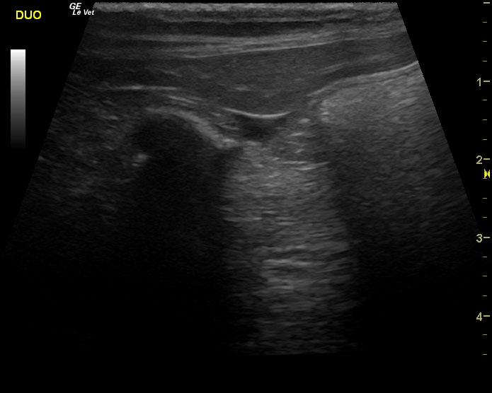
Duodenal shadowing foreign body with concurrent IBD gastrointestinal pattern; neoplasia (lymphoma, mast cell), FIP.
Image Interpretation
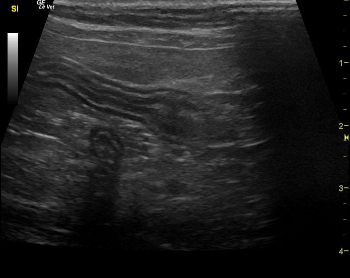
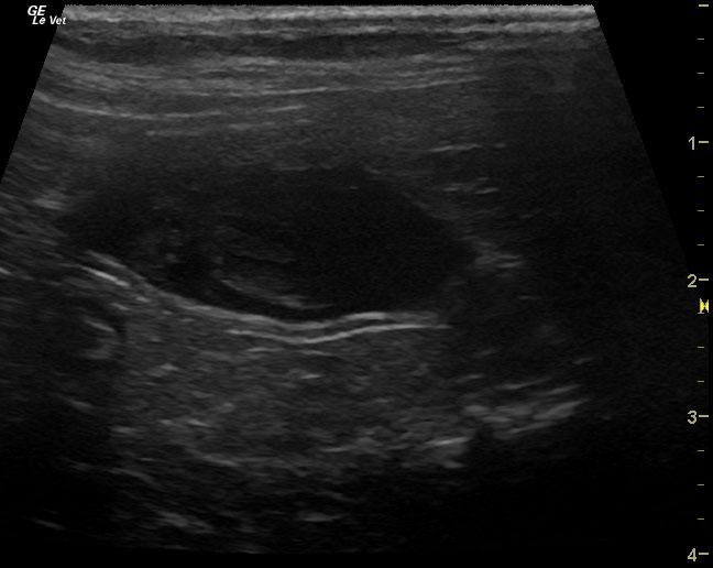
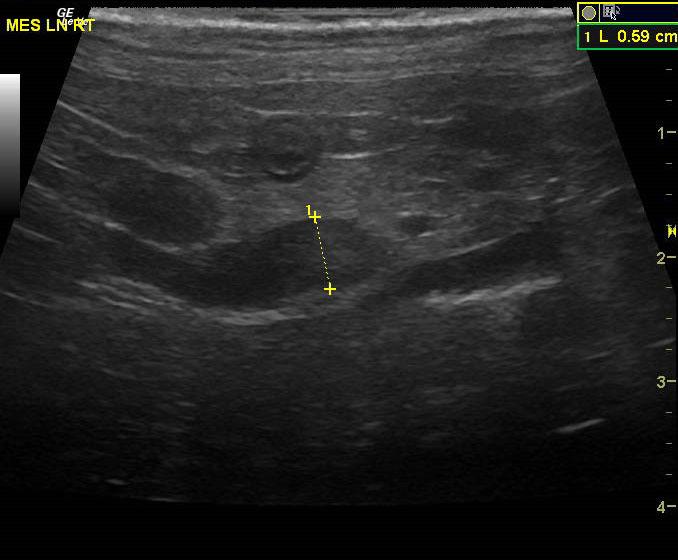
The gastrointestinal tract presented variable areas of intestinal wall thickening with mesenteric root lymphadenopathy that measured 0.6 x 1.2 cm and was uniform and longer than it was wide. Therefore, it would be most suggestive of lymphadenitis. Minor areas of loss of intestinal wall detail was noted. The mid jejunum was thickened with loss of detail. The duodenum revealed echogenic shadowing material that measured 1.5 cm with an additional sharp object that measured 0.7 cm. The ileocecal valve appeared free of evident pathology with a normal dirty shadow in the cecum.
DX
Gastrointestinal foreign body
Outcome
Surgery was performed and foreign bodies (small charcoal pieces or possibly potpourri pieces) were removed from 2 areas. Cat has put on 1 pound since surgery.
Comments
It is possible that the foreign shadowing structures may represent medications that are not dissolved in the stomach given the size and echogenic shadowing. However, it is more suggestive of foreign bodies. Complete evaluation of oral medications given to this patient in the last 48 hours should be evaluated. Exploratory surgery with enterotomy and likely milk through the upper duodenal foreign matter would be recommended with concurrent gastrointestinal biopsies. It is possible the foreign bodies move distally between the time of the ultrasound and surgery.
Clinical Differential Diagnosis
vomiting and anorexia: GI pathology ( neoplasia, ulceration, foreign body, IBD, abscessation/granulomatous disease; pancreatic pathology ( pancreatitis, neoplasia)
Sampling
none
Video
Patient Information
Patient Name :
Rogue R
Gender :
Male, Neutered
Species :
Feline
Type of Imaging : Ultrasound
Status :
Complete
Liz Wuz Here :
Yes
Code :
04_00280
Clinical Signs
- Anorexia
- Vomiting
Exam Finding
- Palpable mass
Images
Blood Chemistry
- Total Protein, Low
CBC
- WBC, High
- WBC, Low
Clinical Signs
- Anorexia
- Vomiting
Urinalysi
- Calcium Oxalate Crystals Present