This 13-year-old FS DLH cat was presented for occasional vomiting over the last 1.5 years. Clinical exam and blood analysis were unremarkable.
This 13-year-old FS DLH cat was presented for occasional vomiting over the last 1.5 years. Clinical exam and blood analysis were unremarkable.
Case Study
Gastric Trichobezoar in a 13 year old FS DLH cat
Sonographic Differential Diagnosis
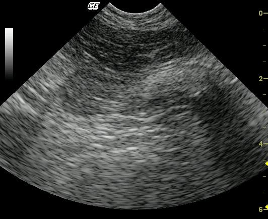
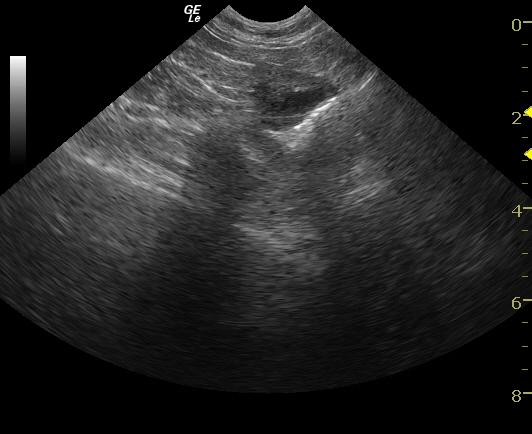
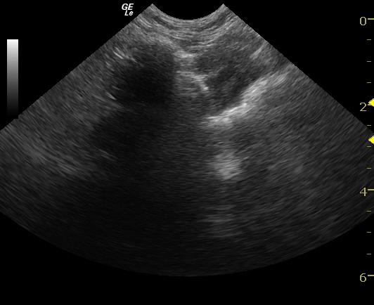
Suspect partially or intermittently obstructing, minimally dense foreign gastric material (i.e. hairball, cloth, paper).
Image Interpretation
The pylorus is moderately distended with echogenic material with dirty distal acoustic shadowing. On the longitudinal view, the material is ovoid to fusiform.
DX
Tricobezoar. Lymphoplasmacytic gastritis with helicobacter.
Outcome
The patient responded well to surgery and still occasionally vomits but is responsive to treatment for inflammatory bowel disease and helicobacter infection.
Comments
No further outcome.
Clinical Differential Diagnosis
GI pathology – Chronic trichobezoar, gastroenteritis, IBD, dietary intolerance, Helicobacter, neoplasia; pancreatic pathology- pancreatitis; hyperthyroidism, .
Sampling
Surgical biopsies revealed moderate lymphoplasmacytic gastritis with helicobacter infection. A large trichobezoar was removed from the gastric fundus. The changes were localized within the mucosa with no evident pathology in the submucosa or muscularis.
Video
Patient Information
Patient Name :
Kitty R
Gender :
Female, Spayed
Species :
Feline
Type of Imaging : Ultrasound
Status :
Complete
Liz Wuz Here :
Yes
Code :
04_00091
Clinical Signs
- Vomiting
Images
Clinical Signs
- Vomiting