A 7-year-old MN Dachshund was presented for anorexia, dehydration, and vomiting for two days. Recent CBC and chemistry panel had shown elevated HCT, neutrophilia, monocytosis, low sodium, low potassium and low chloride, and elevated ALP; elevated AST activity, elevated amylase, increased anion gap, increased bicarbonate, elevated urea, elevated cholesterol, elevated creatinine, elevated phosphate and elevated CK. 4DX snap ELISA was negative and urinalysis and fecal examination within normal limits.
A 7-year-old MN Dachshund was presented for anorexia, dehydration, and vomiting for two days. Recent CBC and chemistry panel had shown elevated HCT, neutrophilia, monocytosis, low sodium, low potassium and low chloride, and elevated ALP; elevated AST activity, elevated amylase, increased anion gap, increased bicarbonate, elevated urea, elevated cholesterol, elevated creatinine, elevated phosphate and elevated CK. 4DX snap ELISA was negative and urinalysis and fecal examination within normal limits.
Case Study
Intestinal foreign body in a 7 year old MN Dachshund dog
Sonographic Differential Diagnosis
Two separate foreign bodies with obstructive pattern of the ascending duodenum and jejunum. Reactive steatitis. Potential for emerging peritonitis.
Image Interpretation
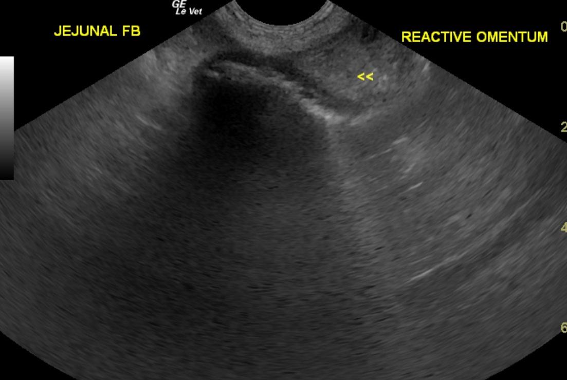
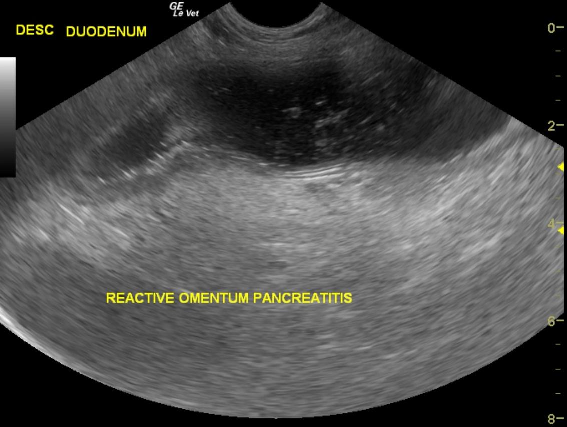
The stomach in this patient presented a significant amount of dilation with thickened pylorus and irregular mucosal tissue. The duodenum was severely dilated with echogenic omentum and mild to moderate hyperechoic pancreatic changes along the right limb. The duodenum was hyperperistaltic and filled with fluid to the duodenal flexure. In the ascending duodenum this reached a cloth type foreign body obstructing the duodenal outflow. This was followed by empty small intestine, which in turn was followed by a second jejunal foreign body that appeared fluid absorbing without overt obstruction. However, some reactive omentum was associated with the intestines. The surgeon should expect multiple areas of potential necrotic intestine and need for resection. No free fluid was noted in the abdomen at this time. Mild to moderate degenerative renal changes.
DX
Outcome
The patient was admitted to an emergency referral hospital where on exploratory laparotomy a peach pit was removed.The patient recovered from surgery without event. At “sutures out” appointment the patient was found to be doing well with a healed incision.
Clinical Differential Diagnosis
GI pathology- foreign body obstruction, neoplasia, partial torsion, intestinal perforation. Peritonitis. pancreatic pathology – pancreatitis; Addison’s disease; liver pathology – toxins, infection.
Sampling
not done
Video
Patient Information
Clinical Signs
- Anorexia
- Dehydration
- Vomiting
Exam Finding
- Dehydration
Images
Blood Chemistry
- Alkaline Phosphatase (SAP), High
- ALT (SGPT), High
- Amylase, High
- Anion Gap, High
- AST (SGOT), High
- Chloride, Low
- Cholesterol, High
- CPK, High
- Creatinine, High
- Phosphorus, High
- Potassium, Low
- Sodium, Low
CBC
- Hematocrit, High
- Monocytes, High
- Neutrophils, High
Clinical Signs
- Anorexia
- Dehydration
- Vomiting
Special Testing
- 4Dx Negative