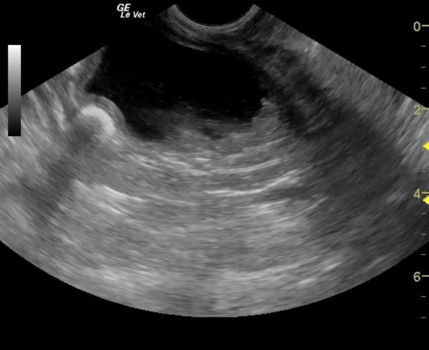
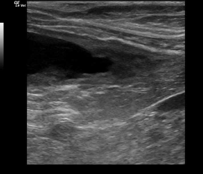
An 11-year-old spayed female German Shepherd dog was presented for leaking urine. On physical examination, the patient was quiet, alert and responsive, and had matted, wet hair on the vulva. Abnormalities on urinalysis included proteinuria, 3+ hematuria, leukocyturia, cocci bacteria, a few triple phosphate and amorphous urate crystals. Urine culture yielded coagulase positive Staphylococcus spp. Urine microalbumin was markedly elevated. The patient was treated with Clavamox, but was presented several days later for ongoing leaking of urine.
An 11-year-old spayed female German Shepherd dog was presented for leaking urine. On physical examination, the patient was quiet, alert and responsive, and had matted, wet hair on the vulva. Abnormalities on urinalysis included proteinuria, 3+ hematuria, leukocyturia, cocci bacteria, a few triple phosphate and amorphous urate crystals. Urine culture yielded coagulase positive Staphylococcus spp. Urine microalbumin was markedly elevated. The patient was treated with Clavamox, but was presented several days later for ongoing leaking of urine. Physical examination and survey abdominal radiographs were both within normal limits. The patient was treated with Simplicef and Rimadyl.