A 9-year-old FS mixed breed dog was presented for weight loss, decreased appetite, and licking of her paws. Physical exam was unremarkable. Thrombocytosis was present on the CBC. Blood chemistry showed an increased urea, hyperproteinemia, hyperglobulinemia, hyperphosphatemia, and elevations of the ALT and elevated GGT enzyme activities.
A 9-year-old FS mixed breed dog was presented for weight loss, decreased appetite, and licking of her paws. Physical exam was unremarkable. Thrombocytosis was present on the CBC. Blood chemistry showed an increased urea, hyperproteinemia, hyperglobulinemia, hyperphosphatemia, and elevations of the ALT and elevated GGT enzyme activities.
Case Study
03_00165 Ruby K Hepatocellular carcinoma
Sonographic Differential Diagnosis
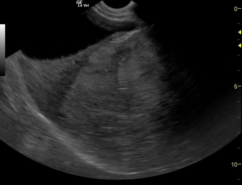
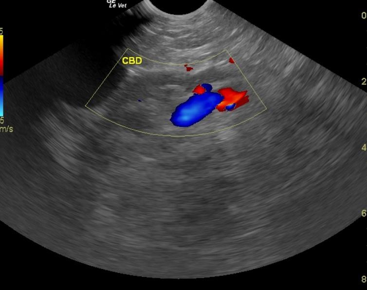
6-4: Persistently large right medial liver mass.No significant progression. Minor portal hypertension. 10-27: Progressive hepatic mass with gastric, common bile duct, and portal deviation. Significant progression. Poor long term prognosis.
Image Interpretation
6-4: The hepatic mass in this patient is persistently micro and macrocystic which now presents with periportal lymphadenopathy. The size is about the same as it was approximately 8 months ago, however impingement upon the portal vein is causing minor congestion into the portal vein and resulting in minor splenomegaly and passive congestion. However this is likely not clinically significant at this time. Other minor micronodular changes were noted in the left liver. 10-27: The hepatic mass in this patient is significantly expanded caudally and medially deviating the portal vein and common bile duct expanding upon the gastrointestinal tract causing gastrointestinal deviation caudally. The gallbladder was impinged upon and partially obstructed causing excessive debris accumulation. The remainder of the abdomen did not present any progression.
DX
Hepatocellular carcinoma.
Clinical Differential Diagnosis
Cholangitis with a possible mucocele causing pain, nausea and therefore loss of appetite and weight loss, hepatitis – Chronic active hepatitis, infectious (leptospirosis, toxoplasmosis), immune-mediated with secondary ascending bacterial infection, pancreatitis with secondary cholestasis and/or hepatitis, neoplasia (primary vs. metastatic; hepatocellular adenoma, lymphoma, carcinoma, leiomyoma, leiomyosarcoma, mast cell tumor, histiocytic sarcoma, etc.)
Sampling
U/S- guided Tru-cut liver biopsy.The patient was sent to a referral hospital for a CT scan and biopsy of the liver mass. The CT scan confirmed the ultrasound findings of a non-resectable mass involving the right liver. Results of the biopsy revealed a hepatocellular carcinoma. The owner elected to keep the dog comfortable with the intention to pursue humane euthanasia once he no longer had a good quality of life (i.e. appetite, activity level, weight).
Video
Patient Information
Gender :
Female, Spayed
Species :
Canine
Type of Imaging : Ultrasound
Status :
Complete
Clinical Signs
- Anorexia
- Weight loss
Images
Blood Chemistry
- ALT (SGPT), High
- GGT High
- Globulin, High
- Phosphorus, High
- Total Protein, High
Clinical Signs
- Anorexia
- Weight loss