An 8 year-old FS Beagle dog was presented for intermittent diarrhea of several months duration. There was no history of vomiting, and the dog remained active and continued to eat well. The dog had undergone an exploratory laparotomy in 2000 at which time multiple enlarged intra-abdominal lymph nodes were identified. Biopsy results of the enlarged lymph nodes showed reactive hyperplasia without any signs of neoplasia. Radiographs were performed but showed decreased detail due to the patient’s obesity; however, no obvious abnormalities were noted.
An 8 year-old FS Beagle dog was presented for intermittent diarrhea of several months duration. There was no history of vomiting, and the dog remained active and continued to eat well. The dog had undergone an exploratory laparotomy in 2000 at which time multiple enlarged intra-abdominal lymph nodes were identified. Biopsy results of the enlarged lymph nodes showed reactive hyperplasia without any signs of neoplasia. Radiographs were performed but showed decreased detail due to the patient’s obesity; however, no obvious abnormalities were noted. Blood chemistry revealed hyperkalemia, while the CBC showed a leukocytosis, consisting of a neutrophilia, and monocytosis, as well as a thrombocytosis.
Case Study
Lymphoma in an 8 year old FS Beagle dog
Sonographic Differential Diagnosis
Abdominal infiltrative disease such as lymphoma, primarily involving the distal intestinal tract.
Image Interpretation
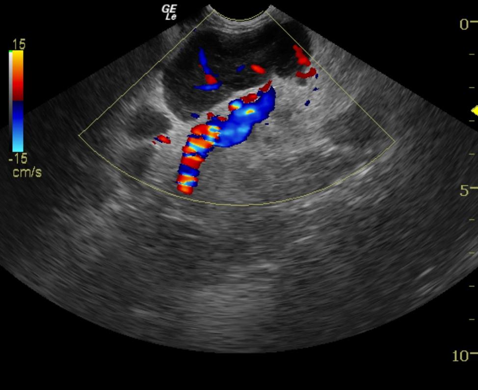
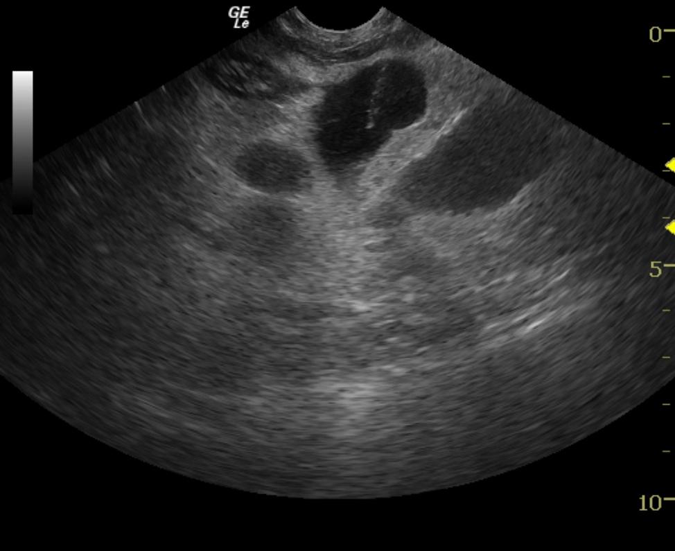
The abdomen in this patient presented multiple mesenteric root lymph nodes that were significantly enlarged and dramatically hypoechoic, significant loss of parenchymal detail and inflamed capsules with reactive surrounding mesentery. The colon was diffusely infiltrated with mural detail loss. The distal small intestine was also infiltrated. The iliac lymph nodes were enlarged with similar presentation of the mesenteric root lymph nodes
DX
Outcome
Chemotherapy was initiated, but the dog became anorexic, adipsic and lost a significant amount of weight. She also developed hematochezia, at which time the owner elected to euthanize the dog for humane reasons.
Clinical Differential Diagnosis
GI pathology (food allergy or intolerance, dietary indiscretion, gastrointestinal parasitism, inflammatory bowel disease, antibiotic responsive diarrhea, gastrointestinal neoplasia -lymphoma, adenocarcinoma, leiomyoma, leiomyosarcoma, mast cell tumor.
Sampling
US-guided FNA of the mesenteric lymph nodes was performed at an angle to avoid the mesenteric artery (CF).
Video
Patient Information
Clinical Signs
- Diarrhea
Images
Blood Chemistry
- Potassium, High
CBC
- Monocytes, High
- Neutrophils, High
- Platelet Count, High
- WBC, High
Clinical Signs
- Diarrhea