A 4-year-old MN Leonberger dog was presented for intermittent vomiting and a decreased appetite. The dog was in thin body condition on physical exam. The CBC showed leukopenia, neutropenia, lymphopenia, thrombocytopenia, moderate anisocytosis, moderate poikilocytosis, and moderate acanthocytosis. cTLI, vitamin B12, and folate were within the normal reference range. The dog was re-evaluated due to persistent vomiting and he was still considered underweight. The spleen and liver could not be delineated on the abdominal radiographs.
A 4-year-old MN Leonberger dog was presented for intermittent vomiting and a decreased appetite. The dog was in thin body condition on physical exam. The CBC showed leukopenia, neutropenia, lymphopenia, thrombocytopenia, moderate anisocytosis, moderate poikilocytosis, and moderate acanthocytosis. cTLI, vitamin B12, and folate were within the normal reference range. The dog was re-evaluated due to persistent vomiting and he was still considered underweight. The spleen and liver could not be delineated on the abdominal radiographs. Urinalysis yielded a cloudy, orange urine with a pH and specific gravity within normal limits, as well as bilirubinuria. The urine culture was negative.
Case Study
Gastric carcinoma in a 4 year old MN Leonberger dog
Sonographic Differential Diagnosis
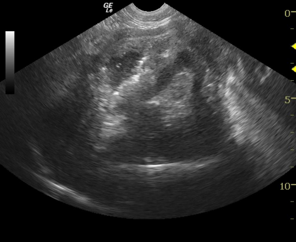
gastric neoplasia such as carcinoma, severe gastritis, potentially ulcerative.
Image Interpretation
The gastric wall is thickened and echogenic with some retained anechoic fluid. Regional loss of mural detail and peristalsis is noted suggestive for neoplasia or aggressive transmural inflammatory disease. The descending duodenum is hyperperistaltic suggestive for enteritis.
DX
Outcome
The owner elected humane euthanasia due to the persistent vomiting and progressive deterioration.
Clinical Differential Diagnosis
Gastrointestinal neoplasia (lymphoma, adenocarcinoma, leiomyoma, leiomyosarcoma, mast cell tumor,) hepatic neoplasia (lymphoma, adenocarcinoma, leiomyosarcoma, histiocytic sarcoma, mast cell tumor,) splenic neoplasia (lymphoma, histiocytic sarcoma, mast cell tumor,) pancreatic neoplasia (adenocarcinoma,)atypical hypoadrenocorticism – less likely given the thrombocytopenia and radiographic abnormalities.
Sampling
US-guided gastric FNA was suggestive for carcinoma. Full thickness biopsies (postmortem) Stomach: transmural, poorly differentiated, gastric carcinoma. Pancreas: no visible microscopic lesion. Myocardium: moderate to severe fatty infiltration (steatitis.)
Video
Patient Information
Clinical Signs
- Anorexia
- Vomiting
Exam Finding
- Weight loss
Images
CBC
- Anisocytosis
- Lymphocytes, Low
- Neutrophils, Low
- Platelet Count, Low
- WBC, Low
Clinical Signs
- Anorexia
- Vomiting
Urinalysi
- Appearance Turbid
- Bilirubin Present
- Color Abnormal