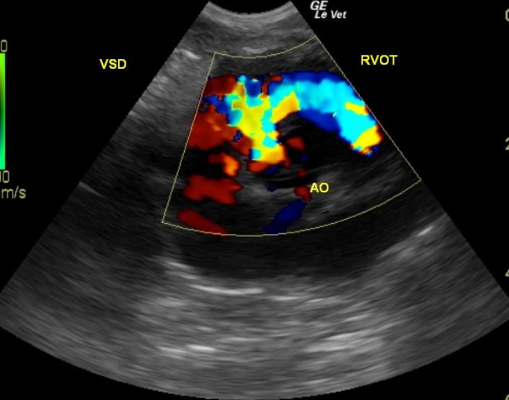
The left ventricle presented normal internal volume with adequate contractility, but the membranous septum presented a defect with a left to right turbulent flow and a velocity of approximately 6 m/s. This defect is most consistent with a ventricular septal defect. The defect was confirmed in a 5 chamber right parasternal long axis (Videos 1-3) and also in right parasternal short axis heart base view (Video 4). Normal chamber sizes and contractility suggest that the defect is clinically compensated at this time. The tricuspid valve was found to be linear with proper extension, length and closure. There was no evidence of tethering, length disparity, or vegetation of the leaflets which would indicate dysplastic morphology. Color flow and pulse wave Doppler assessment of the tricuspid valve revealed adequate laminar flow without hemodynamically significant regurgitation or excessive velocity. The right ventricle demonstrated normal size (1/3 diameter of the left ventricle), morphology, and kinetic activity.