Adrenocortical carcinoma with venous invasion in a 15 year old FS DSH cat

Case Study

Adrenocortical carcinoma with venous invasion in a 15 year old FS DSH cat

A 15-year-old FS DSH with was presented for the evaluation of intermittent vomiting. Blood work performed by the referring veterinarian showed a persistent thrombocytopenia and a progression of the severity of azotemia over a period of several months. A grade I/VI heart murmur was found on physical examination. Blood work was repeated and revealed hypoalbuminemia, hyperamylasemia, hypercalcemia and hypernatremia. The CBC showed a thrombocytopenia and monocytosis. The urinalysis showed a low specific gravity, mild proteinuria, and hematuria.
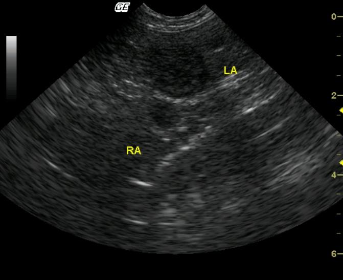
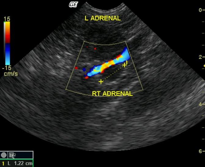
A 15-year-old FS DSH with was presented for the evaluation of intermittent vomiting. Blood work performed by the referring veterinarian showed a persistent thrombocytopenia and a progression of the severity of azotemia over a period of several months. A grade I/VI heart murmur was found on physical examination. Blood work was repeated and revealed hypoalbuminemia, hyperamylasemia, hypercalcemia and hypernatremia. The CBC showed a thrombocytopenia and monocytosis. The urinalysis showed a low specific gravity, mild proteinuria, and hematuria.

Sonographic Differential Diagnosis

There appears to be a left adrenal mass which is wrapping around the aorta, however this could also be a primary mass with adjacent lymphadenopathy. Carcinoma most likely, but a pheochromocytoma is also possible.

Image Interpretation

A large hypoechoic mass is noted in the area of the left adrenal in the near field. A separate hypoechoic round structure is also present in the far field wrapping the aorta. These two structures may be connected or represent a primary adrenal mass with adjacent lymphadenopathy.

DX

Adrenocortical carcinoma with venous invasion.

Outcome

Three radiographic views of the thorax did not show signs of metastases. A left adrenalectomy was performed and complete excision of the mass was achieved. Initially, the patient did well post-operatively, however, she soon developed sunken eyes and extremely pale to white mucous membranes. Supportive care and blood transfusion allowed for clinical stabilization of the patient. Blood chemistry revealed hypoalbuminemia, low ALT, azotemia, hyperglycemia, hypernatremia, and hypokalemia. The CBC showed lymphopenia, monocytosis, neutrophilia, eosinophilia, and basophilia, in addition to a decreased hematocrit, and thrombocytopenia. CBC values slowly improved over the course of about eight weeks. Two months later, the patient was still azotemic with a high ALT, hypernatremia, and hyperamylasemia. CBC was now within normal limits. A consult with an oncologist yielded no effective chemotherapy protocols for this type of cancer. The patient was euthanized almost one year after the tumor had been originally discovered by ultrasound.

Clinical Differential Diagnosis

Hyperaldosteronism secondary to an adrenal mass (carcinoma or adenoma,) pheochromocytoma, protein losing nephropathy due to neoplasia (lymphoma.)

Sampling

The cat was referred for surgical adrenalectomy. Histopathology returned as adrenocortical carcinoma with venous invasion.

Patient Information

Patient Name : Emma B
Gender : Female, Spayed
Species : Feline
Type of Imaging : Ultrasound
Status : Complete
Liz Wuz Here : Yes
Code : 07_00021

Clinical Signs

  • Vomiting

Exam Finding

  • Heart Murmur

Images

LeftadrenalmassColorstill

Blood Chemistry

  • Albumin, Low
  • Amylase, High
  • Azotemia
  • Calcium, High
  • Sodium, High

CBC

  • Monocytes, High
  • Platelet Count, Low

Clinical Signs

  • Vomiting

Urinalysi

  • Blood Present
  • Protein Present
  • Specific Gravity Low