A 6-year-old intact male English Bulldog was presented for 2-3 days of intermittent vomiting and lethargy. Physical exam found the patient to be tachycardic with pale mucous membranes and a palpable mass in the abdomen. Serum biochemistry revealed elevated alkaline phosphatase, hypernatremia, and mild hypochloremia. On CBC, a decreased HCT in conjunction with a high MCV was noted, in addition to a neutrophilia, monocytosis and thrombocytopenia. No abnormalities were noted on thoracic radiographs.
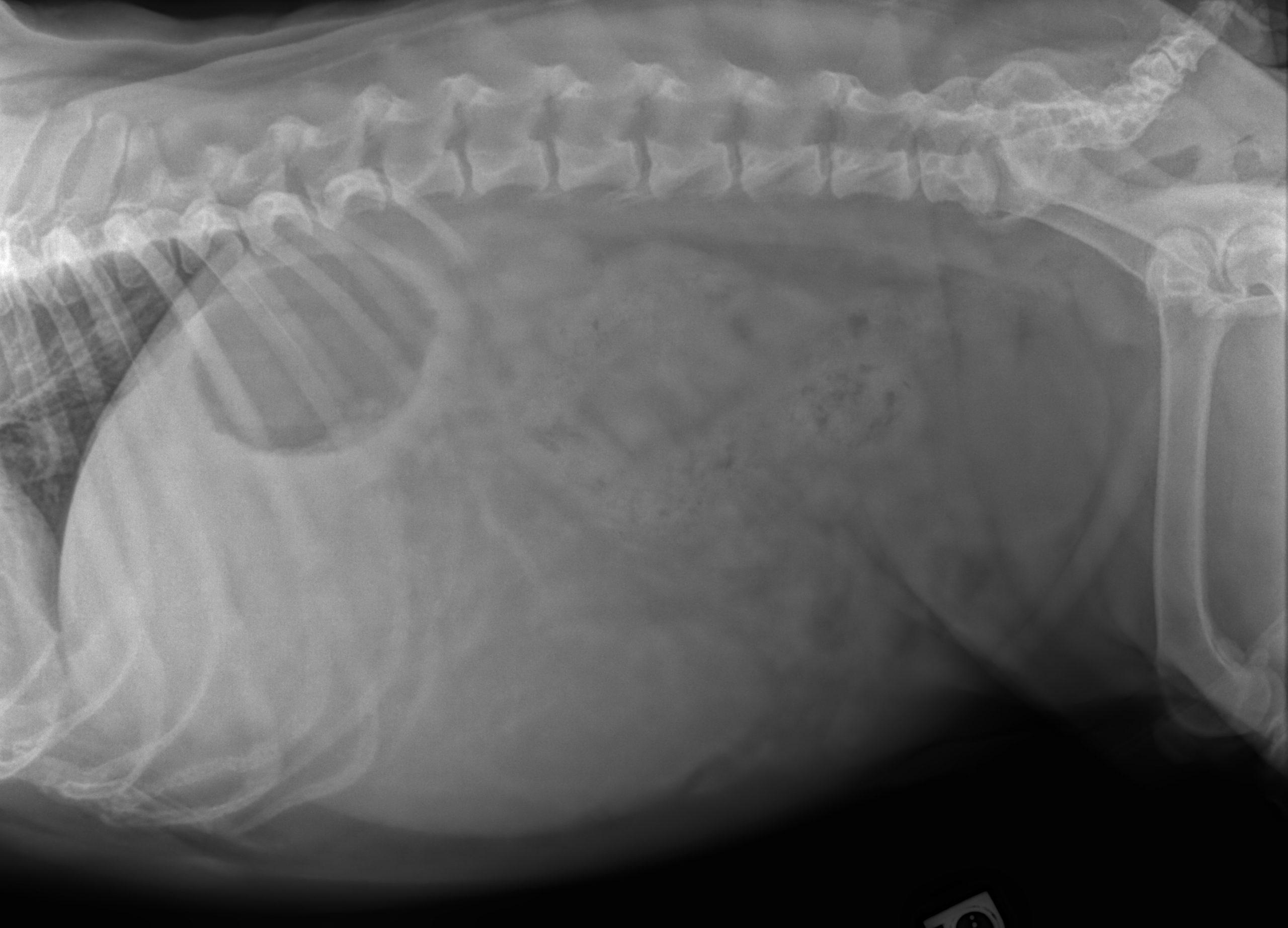
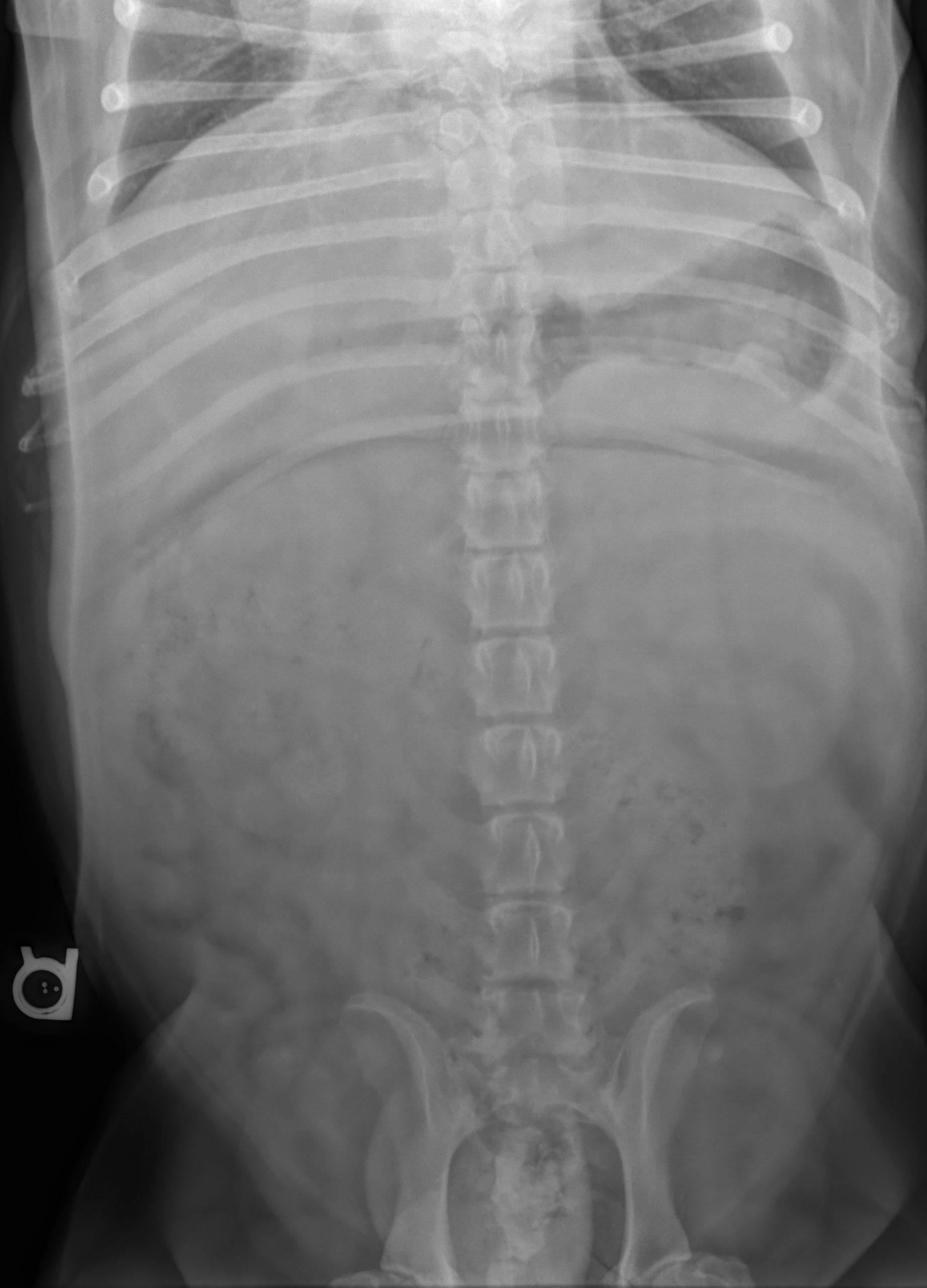
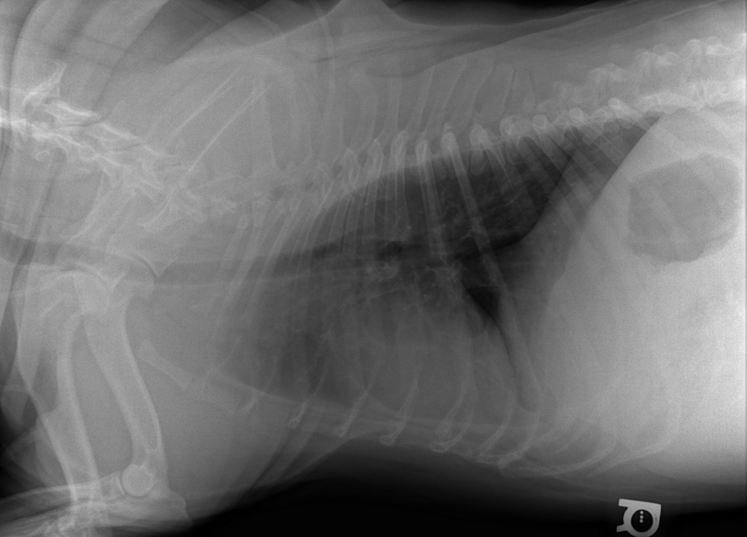
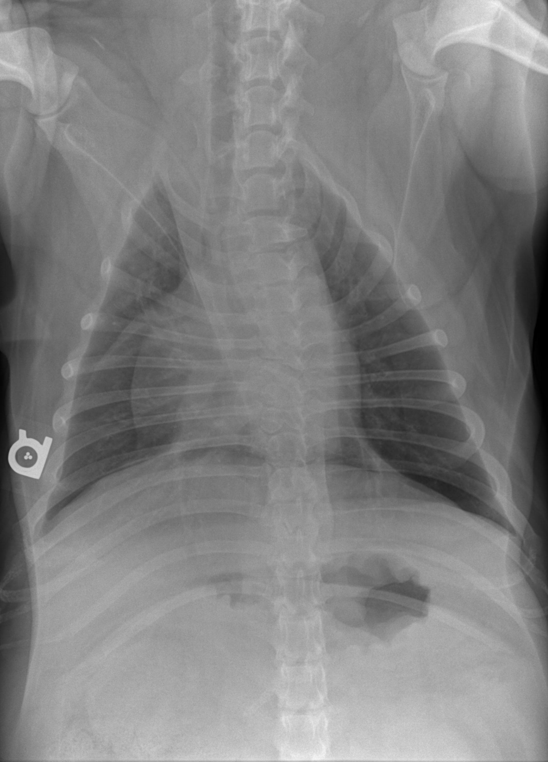
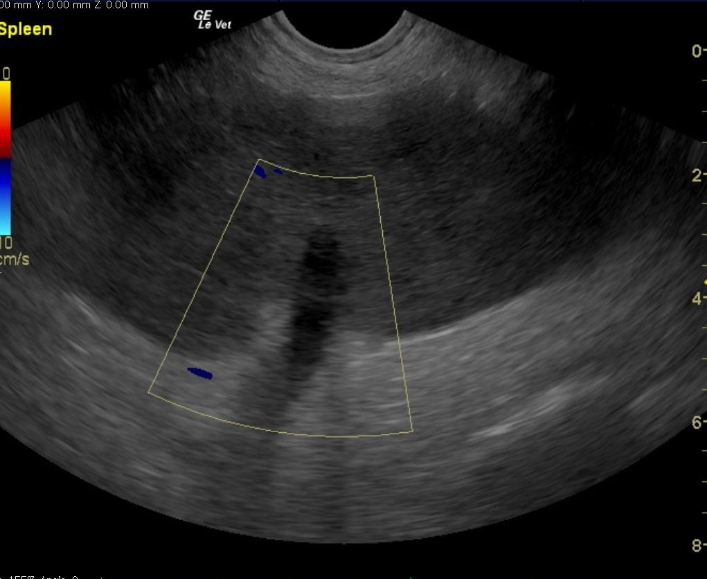
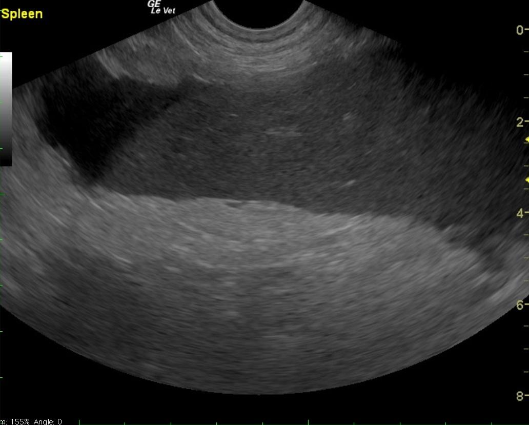
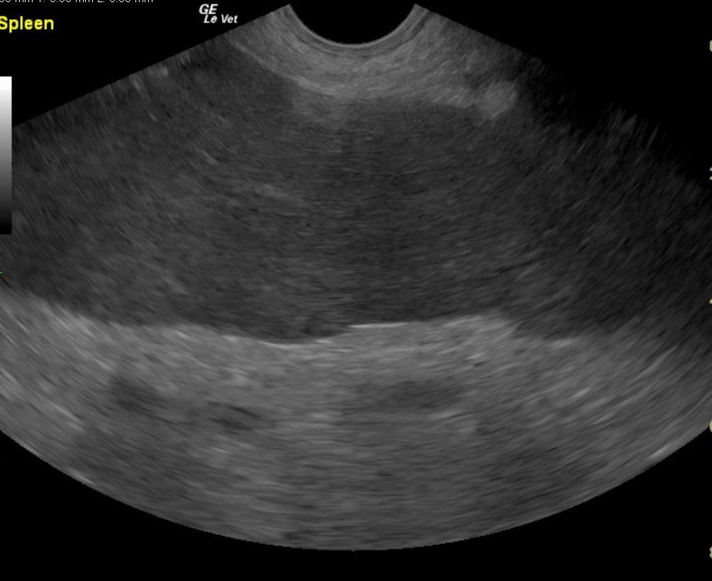
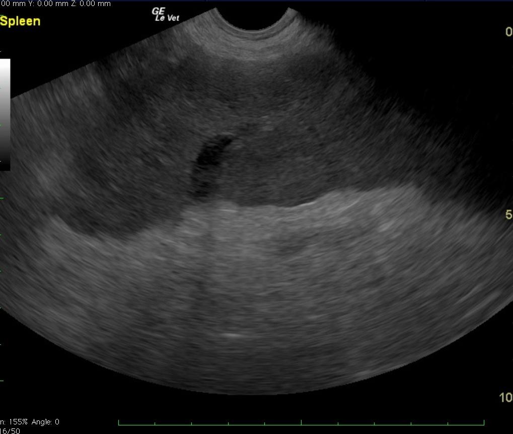
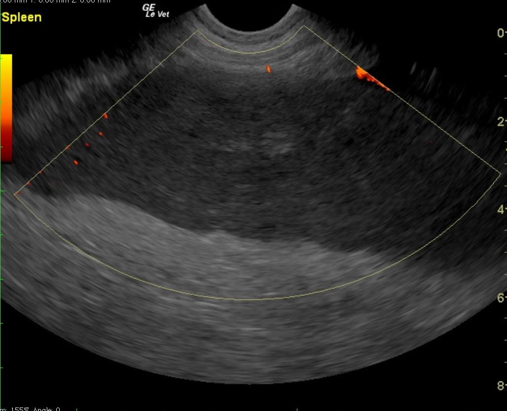
A 6-year-old intact male English Bulldog was presented for 2-3 days of intermittent vomiting and lethargy. Physical exam found the patient to be tachycardic with pale mucous membranes and a palpable mass in the abdomen. Serum biochemistry revealed elevated alkaline phosphatase, hypernatremia, and mild hypochloremia. On CBC, a decreased HCT in conjunction with a high MCV was noted, in addition to a neutrophilia, monocytosis and thrombocytopenia. No abnormalities were noted on thoracic radiographs. Abdominal radiographic findings revealed a loss of contrast in the cranial abdomen, with the possibility of a splenic mass. Radiographic Interpretation showed marked splenomegaly strongly suspected to be secondary to torsion, although accompanying infiltrative or neoplastic pathology could not be ruled out. The small volume of peritoneal effusion and inflammation was likely secondary to the splenic pathology. Thoracic radiographs showed mild microcardia and under perfused pulmonary vasculature in support of hypovolemia. Atypical intrathoracic fat distribution was considered to be incidental in this dog. This dog had multiple hemivertebrae.