A 13-year-old MN mixed breed dog, with history of chronic intermittent vomiting, was presented for progressively worsening vomiting and decreased appetite. Physical exam was unremarkable. 4DX test was positive for Lyme disease and C6 had a clinically significant titer. Several months later, the patient was having increasing frequency of vomiting. The owner reported that the patient had been vomiting bile and food weekly. Blood chemistry revealed hyperlipasemia. The patient was discharged with gastro-protectant medication.
A 13-year-old MN mixed breed dog, with history of chronic intermittent vomiting, was presented for progressively worsening vomiting and decreased appetite. Physical exam was unremarkable. 4DX test was positive for Lyme disease and C6 had a clinically significant titer. Several months later, the patient was having increasing frequency of vomiting. The owner reported that the patient had been vomiting bile and food weekly. Blood chemistry revealed hyperlipasemia. The patient was discharged with gastro-protectant medication. A few weeks later, the patient represented due to continued frequent vomiting and decreased appetite. The patient was administered subcutaneous fluids, gastro-protectant, and antibiotic injections pending abdominal ultrasound.
Case Study
Gastric carcinoma in a 13 year old MN mixed breed dog
Sonographic Differential Diagnosis
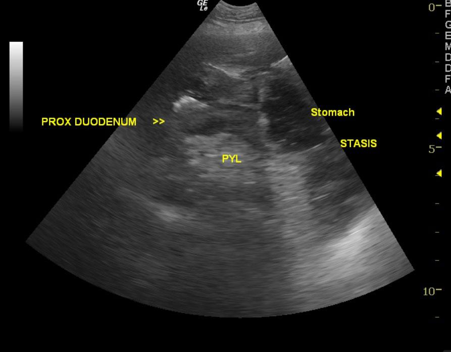
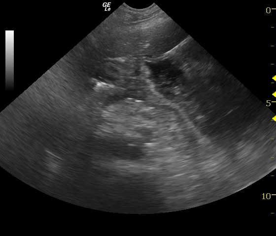
Gastric thickening. (This is likely nonresectable due to position). Neoplastic criteria are present. Carcinoma or round cell neoplasia likely, but there is a potential for ulcerative gastritis or other inflammatory disease.
Image Interpretation
An annular thickening is noted with loss of mural detail in the pyloric antrum. Reactive surrounding fat suggests emerging peritonitis and transmural pathology. (The right intercostal approach is utilized to best visualize the pylorus in this deep chested dog.)
DX
Outcome
The patient underwent exploratory surgery with removal of the mass within the pyloric valve. The patient recovered from anesthesia without complication. Results from the biopsy of the mass revealed gastric carcinoma, and a guarded prognosis was given. At sutures out, physical exam found non-painful abdomen and dog looking well. The patient was sent for evaluation at an oncologist; chemotherapy options were discussed, but owner declined treatment. At a recheck exam approximately 1 month later, the owner reported that the patient had a decreased appetite, was eating primarily table food, was lethargic, and was vomiting dark liquid every two days. The patient was given a steroid injection and sent home with anti-emetic medication. The patient became anorexic and vomiting continued. The owner opted to take patient home for the evening and euthanize the next day.
Clinical Differential Diagnosis
Gastroenteritis, IBD, pyloric outflow obstruction, pancreatitis, gastrointestinal foreign body, neoplasia- gastrointestinal, pancreatic disease.
Sampling
Full-thickness surgical biopsy of the mass revealed gastric carcinoma.
Video
Patient Information
Clinical Signs
- Anorexia
- Vomiting
Images
Blood Chemistry
- Lipase, High
Clinical Signs
- Anorexia
- Vomiting
Special Testing
- 4Dx Positive
- Lyme Positive