An 8-year-old FS Beagle dog was presented for lethargy, anorexia, and vomiting. Blood chemistry (9/23/04) revealed elevated Alkaline Phosphatase, high ALT, high CK, hypercholesterolemia, and high anion gap. CBC was unremarkable. T4 was within normal range. Urine Cortisol/Creatinine level (10/11/05) was high. A low-dose dexamethasone suppression test (10/19/05) revealed hyperadrenocorticism.
An 8-year-old FS Beagle dog was presented for lethargy, anorexia, and vomiting. Blood chemistry (9/23/04) revealed elevated Alkaline Phosphatase, high ALT, high CK, hypercholesterolemia, and high anion gap. CBC was unremarkable. T4 was within normal range. Urine Cortisol/Creatinine level (10/11/05) was high. A low-dose dexamethasone suppression test (10/19/05) revealed hyperadrenocorticism.
Case Study
Hepatocellular carcinoma and gallbladder mucocele in a 8 year old FS Beagle dog
Sonographic Differential Diagnosis
Gall bladder mucocele. Left medial liver mass appears resectable. Likely carcinoma, potential for dramatic nodular hyperplasia/hepatoma. Guarded prognosis.
Image Interpretation
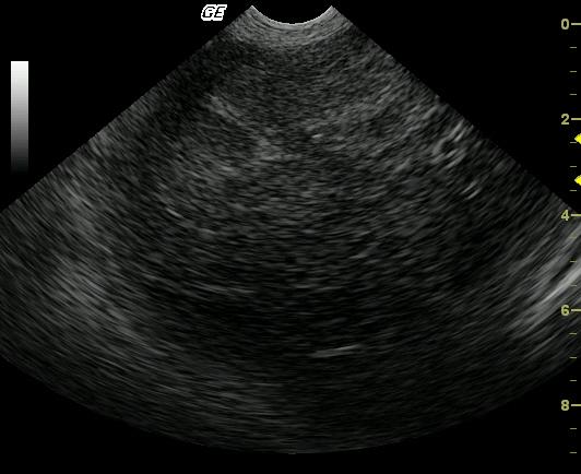
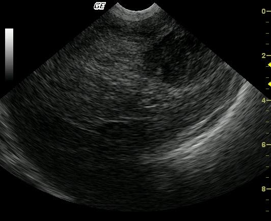
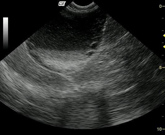
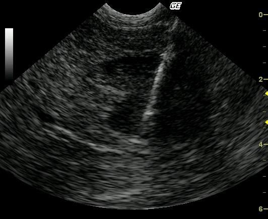
The liver presented a 4cm hypoechoic left medial mass that extended into a larger hyperechoic lesion. Together they measured approximately 8cm. This was located adjacent to the pylorus and approximately 1.5-2cm left of the portal vein. The right liver appeared unremarkable. No periportal lymphadenopathy was noted. The gallbladder was significantly distended with suspended debris and stellate type appearance with mildly ill defined wall contour most consistent with gallbladder mucocele. This was considered moderate. Concurrent cholecystitis is suspected. The biopsy of the liver mass was performed 18 months post cholecystectomy and surgical biopsy was benign (see below).
DX
Outcome
Patient underwent exploratory/cholecystectomy surgery (11/26/06) at referral facility with repeat biopsy of liver mass and culture and sensitivity of gallbladder. Patient was discharged the next day with antibiotics and pain medication. In-house chemistry (1/3/06) revealed high ALT, hypercholesterolemia, slight hyperglycemia, and hyperphosphatemia.
Comments
The patient was lost to follow up after the liver mass was diagnosed as carcinoma.
Clinical Differential Diagnosis
Hyperadrenocorticism: pituitary dependent vs. adrenal neoplasia; Liver pathology: hepatitis, hepatopathy-toxic, metabolic, biliary disease (gallbladder mucocele, choleliths, cholecystitis), neoplasia
Sampling
Gb mucocele was surgically removed along with biopsy of the mass. Culture of gallbladder yielded scant growth of Staphylococcus intermedius. Results from liver mass biopsy (01/04/06) revealed necrosis with fibrosis, mild chronic inflammation, mucoid material accumulation and steroid hepatopathy. 18 months later liver mass biopsy revealed hepatocellular carcinoma (Biopsy video). A 16 gauge ultrasound guided biopsy was performed of the left medial liver mass. The more hypoechoic region of the mass was sampled as it appears to be the more aggressive portion of the lesion.
Video
Patient Information
Clinical Signs
- Anorexia
- Lethargy
- Vomiting
Images
Blood Chemistry
- Alkaline Phosphatase (SAP), High
- ALT (SGPT), High
- Anion Gap, High
- AST (SGOT), High
- Cholesterol, High
- CPK, High
Clinical Signs
- Anorexia
- Lethargy
- Vomiting
Special Testing
- LDDST Positive for Cushing's
Urinalysi
- Urine Cortisol/Creatinine, High