A 12-year-old MN German Shepherd Dog was presented for persistent vomiting and anorexia over a 3 day period. The physical exam revealed moderate dehydration and a palpably enlarged and painful spleen. The CBC presented slight neutropenia. Blood chemistry analysis revealed only a moderately elevated amylase.
A 12-year-old MN German Shepherd Dog was presented for persistent vomiting and anorexia over a 3 day period. The physical exam revealed moderate dehydration and a palpably enlarged and painful spleen. The CBC presented slight neutropenia. Blood chemistry analysis revealed only a moderately elevated amylase.
Case Study
Intestinal foreign body in a 12 year old MN German Shepherd Dog
Sonographic Differential Diagnosis
Intestinal foreign body. Gastroenteritis. Irregular splenic position.
Image Interpretation
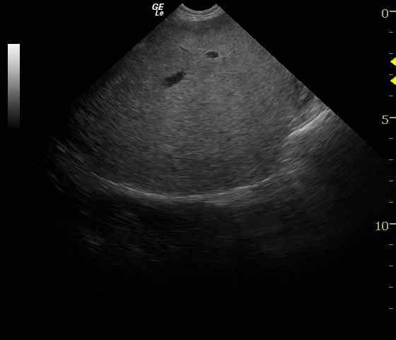
The small intestine presented a shadowing luminal structure consistent with foreign body. Some bunching of small intestine was present with considerable artifact (Video 1). Gastric stasis was present but minimal. The remainder of the intestinal tract appeared edematous most consistent with enteritis. Hyper-peristaltic and dilated small intestine is present with concurrent empty small intestine consistent with an obstructive pattern (Video 2). The spleen was inverted in position and rotated consistent with partial splenic torsion. Vascular congestion was evident (Video 3).
DX
Intestinal foreign body
Outcome
The patient recovered after 3 days of hospitalization and was thriving 20 days post diagnosis.
Comments
See our research from ECVIM 2009 regarding the sonographic criteria for gastrointestinal obstruction in dogs (http://www.sonopath.com/sites/default/files/downloads/Abstract_Highlight_01.pdf).
Clinical Differential Diagnosis
Pancreatitis, gastroenteritis, foreign body, infectious/tick borne disease, splenitis, splenic torsion, breed associated hypersplenism, neoplasia, toxin.
Sampling
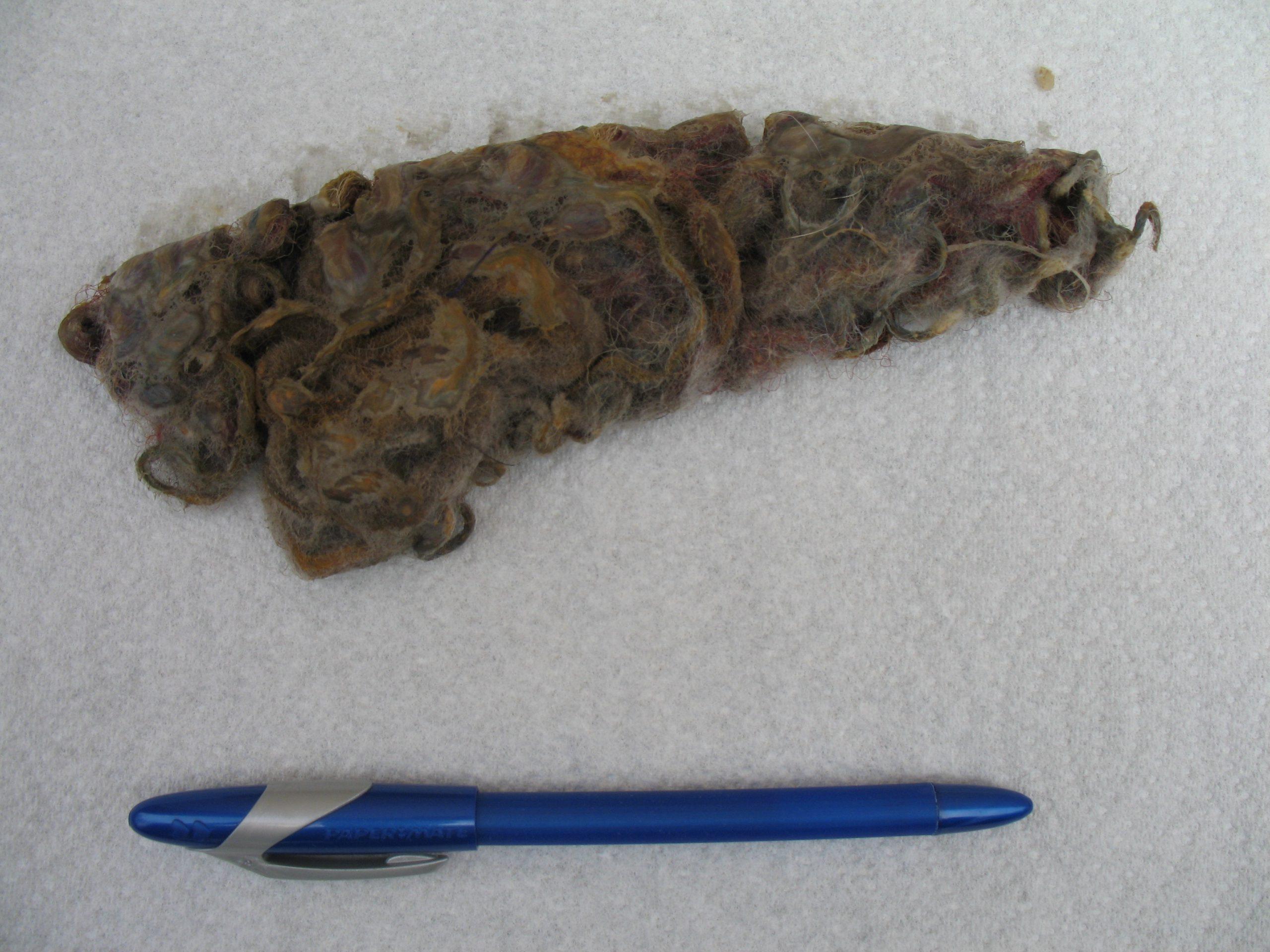
Exploratory surgery revealed a thickened, irregular spleen with no evidence of rupture. Splenectomy was performed. Histopathology of the spleen revealed extensive areas of hemorrhage and hematoma formation, hemosiderosis, extramedullary hematopoiesis, and siderotic capsular plaques. In the intestines, a string foreign body was found and removed, with concurrent resection and anastomosis of small intestine. Histopathology revealed acute on chronic inflammatory bowel.
Video
Patient Information
Patient Name :
Angel G
Gender :
Male, Neutered
Species :
Canine
Type of Imaging : Ultrasound
Status :
Complete
Liz Wuz Here :
Yes
Code :
04_00136
Clinical Signs
- Anorexia
- Vomiting
Exam Finding
- Abdominal Pain
- Dehydration
- Splenomegaly
Images
Blood Chemistry
- Amylase, High
CBC
- Neutrophils, Low
Clinical Signs
- Anorexia
- Vomiting