This 9-year-old MN Boxer was presented for retching, hyporexia, and foul halitosis. The physical exam, CBC, blood chemistry, and urinalyses were all normal. The dog presented 14 hours postprandial of 1 can of i/d diet. The patient had been empirically treated with amoxicillin and metronidazole for presumed Helicobacter gastritis with partial response over a 3 week protocol.
This 9-year-old MN Boxer was presented for retching, hyporexia, and foul halitosis. The physical exam, CBC, blood chemistry, and urinalyses were all normal. The dog presented 14 hours postprandial of 1 can of i/d diet. The patient had been empirically treated with amoxicillin and metronidazole for presumed Helicobacter gastritis with partial response over a 3 week protocol.
Case Study
Delayed Pyloric Emptying in a 9 year old MN Boxer dog
Sonographic Differential Diagnosis
Gastric stasis. Potential causes include a primary motility disorder, mild or emerging inflammatory or infiltrative gastric or perigastric (i.e. pancreatitis) disease in a partially or intermittently obstructing, non-visualized gastric foreign body.
Image Interpretation
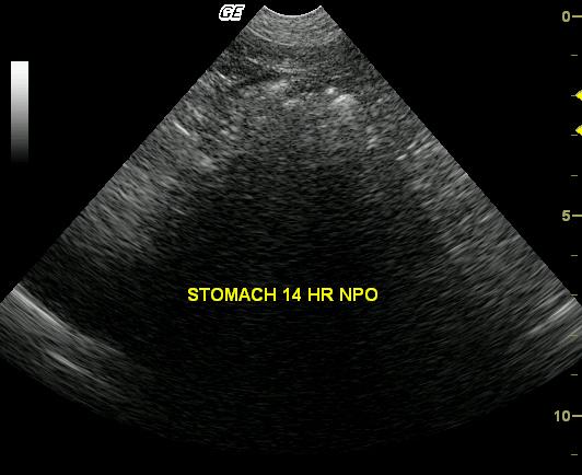
The stomach is moderately filled with echogenic, faintly shadowing ingesta. Pyloric outflow obstruction was not apparent. Luminal distention is hindering critical assessment of gastric mural thickness; however, the discernible wall appears to be within normal limits.
DX
Outcome
This patient presented with simple delayed pyloric emptying secondary to the inflammatory pathology. Endoscopy was selected in this case due to the lack of significant mural pathology that would necessitate full thickness biopsies. The patient responded well to a 6 week treatment for Helicobacter as well as a hypoallergenic diet.
Clinical Differential Diagnosis
GI pathology- Gastric foreign body, gastritis, IBD, neoplasia, pancreatitis.
Sampling
Endoscopic exam after an additional 12 hours NPO revealed an empty lumen with gastric and duodenal mucosal biopsies revealing mild multifocal lymphocytic plasmacytic infiltrate with helicobacter infection.
Video
Patient Information
Clinical Signs
- Anorexia
- Gagging
- Halitosis
Images
Clinical Signs
- Anorexia
- Gagging
- Halitosis