This 8-year-old MN DSH was presented for progressive vomiting and anorexia. The physical exam revealed a palpable cranial abdominal mass, poor body condition, and mild dehydration. CBC presented a mild regenerative anemia while the blood chemistry analysis was normal.
This 8-year-old MN DSH was presented for progressive vomiting and anorexia. The physical exam revealed a palpable cranial abdominal mass, poor body condition, and mild dehydration. CBC presented a mild regenerative anemia while the blood chemistry analysis was normal.
Case Study
Lymphoblastic Lymphoma in an 8 year old MN DSH cat
Sonographic Differential Diagnosis
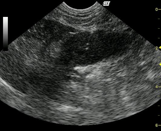
Gastric mural mass compatible with neoplasia.(Lymphoma, round cell tumor such as mast cell) .
Image Interpretation
This cross-sectional image at the gastric fundus reveals marked focal mural thickening with complete loss of wall layering. The gastric wall thickening is adjacent to the liver and in the near field, supporting lesser curvature involvement. The fragmented linear echogenicity noted within the mid aspect of the mass represents a 22 gauge needle.
DX
Lymphoblastic lymphoma
Outcome
The patient initially responded well to a CHOP chemotherapy protocol. However, the owner elected humane euthanasia 10 weeks post diagnosis due to lack of response to therapy.
Clinical Differential Diagnosis
GI pathology – Gastroenteritis, IBD, pancreatitis, neoplasia, gastrointestinal foreign body, trichobezoar, maldigestion.
Sampling
22-gauge US guided FNA revealed lymphoblastic lymphoma.
Video
Patient Information
Patient Name :
Five T
Gender :
Male, Neutered
Species :
Feline
Type of Imaging : Ultrasound
Status :
Complete
Liz Wuz Here :
Yes
Code :
04_00104
Clinical Signs
- Anorexia
- Vomiting
Exam Finding
- Dehydration
- Palpable mass
- Weight loss
Images
CBC
- RBC, Low
Clinical Signs
- Anorexia
- Vomiting