This 6-month-old MI mixed breed dog presented for acute vomiting and poor appetite but otherwise normal. The physical exam was relatively unremarkable except for excessive gastrointestinal activity, borborygmus and moderate hyperthermia.
This 6-month-old MI mixed breed dog presented for acute vomiting and poor appetite but otherwise normal. The physical exam was relatively unremarkable except for excessive gastrointestinal activity, borborygmus and moderate hyperthermia.
Case Study
Ileocecocolic Junction Foreign Body in a 6 month old MI mixed breed dog
Sonographic Differential Diagnosis
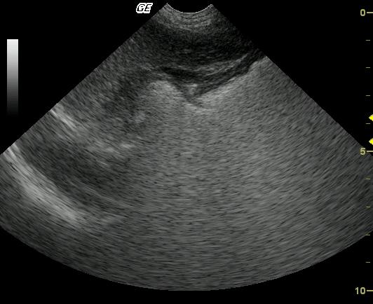
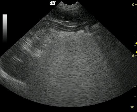
Suspect mucosal sloughing and regional lymphadenopathy consistent with inflammatory disease. The lymph node characteristics support reactive lymphadenopathy (Video 3.) Adhered intraluminal material suggests the presence of blood clots. Primary consideration is given to gastritis secondary to dietary indiscretion with reactive lymphadenitis
Image Interpretation
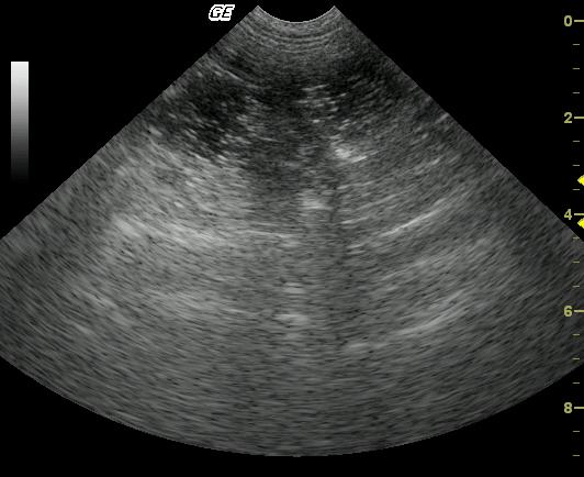
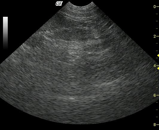
The stomach is mildly to moderately distended with fluid, gas and echogenic ingesta. Discrete, irregularly shaped, hypoechoic, homogeneous structures are also noted within the gastric lumen and may be adhered to the gastric wall, given their persistence in location. The stomach wall is diffusely, moderately thick walled and abnormally layered, represented by a prominent muscularis. Regional thinning of the mucosal layer and is also noted. A mildly to moderately enlarged, normally shaped, and mildly hypoechoic regional lymph node is also present Video 3.Diffuse, moderate gastric wall thickening with stasis. A distal small intestinal stasis pattern was also noted followed by a progressively shadowing luminal structure and empty small intestine (Videos 1 & 2.)
DX
Ileocecocolic junction foreign body
Outcome
The patient thrived postoperatively with IV support, intravenous antibiotics and GI protectant therapy.
Clinical Differential Diagnosis
GI pathology – Dietary indiscretion, GI foreign body, parasitic disease/other infectious, intussusception; pancreatic pathology – pancreatitis; toxicosis.
Sampling
Exploratory laparotomy was performed that revealed an accumulation of sticks, pine needles, hair, and string at the ileocecal junction. Blood clots were present in the stomach. Full thickness gastrointestinal biopsies revealed mild, diffuse, chronic, lymphocytic bacterial gastritis consistent with helicobacter infection. Intestinal biopsies at the ileum revealed moderate lymphoplasmacytic enteritis with Peyer’s Patch hyperplasia. Mesenteric lymph node biopsies revealed moderate eosinophilic lymphadenitis and lymphoid hyperplasia.
Video
Patient Information
Patient Name :
Dunkin P
Gender :
Male, Intact
Species :
Canine
Type of Imaging : Ultrasound
Status :
Complete
Liz Wuz Here :
Yes
Code :
04_00092
Clinical Signs
- Anorexia
- Vomiting
Exam Finding
- Fever
Images
Clinical Signs
- Anorexia
- Vomiting