This 5-year-old FS Bichon Frise dog presented for 5 weeks of vomiting and diarrhea with a recent onset of lethargy. The physical exam revealed mild dehydration and focally painful abdomen. Blood analysis demonstrated mild leukocytosis with a left shift and mild hypoalbuminemia.
This 5-year-old FS Bichon Frise dog presented for 5 weeks of vomiting and diarrhea with a recent onset of lethargy. The physical exam revealed mild dehydration and focally painful abdomen. Blood analysis demonstrated mild leukocytosis with a left shift and mild hypoalbuminemia.
Case Study
Duodenal Foreign Body, Moderate Lymphoplasmacytic Enteritis, in a 5 year old FS Bichon Frise dog
Sonographic Differential Diagnosis
Partially obstructing small bowel foreign body with segmental corrugation with adjacent peritoneal inflammation.
Image Interpretation
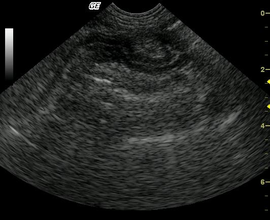
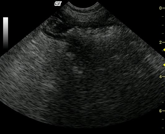
The first image demonstrates a moderately corrugated segment of small bowel with a slightly tortuous appearance and the omentum and mesentery surrounding the small bowel are moderately echogenic. In the next image, a second segment of small bowel is focally, moderately distended with echogenic, shadowing foreign material. The distal small bowel is normally empty and contracted with normal mural architecture.The tortuous small bowel appearance with adjacent peritoneal inflammation is often seen with adhesions which are strongly suspected in this case
DX
Duodenal foreign body, moderate lymphoplasmacytic enteritis, focal eosinophilic imflammation
Outcome
The patient responded well postoperatively with resolution of symptoms at a 3-month follow-up exam. Albumin levels returned to normal. Medical treatment included a hypoallergenic diet with supportive antibiotics. The patient was clinically normal 2 years after the original presentation.
Clinical Differential Diagnosis
GI pathology – gastroenteritis/IBD pancreatitis, foreign body, intestinal ulceration/perforation/peritonitis, bowel infarction, neoplasia; Addison`s disease
Sampling
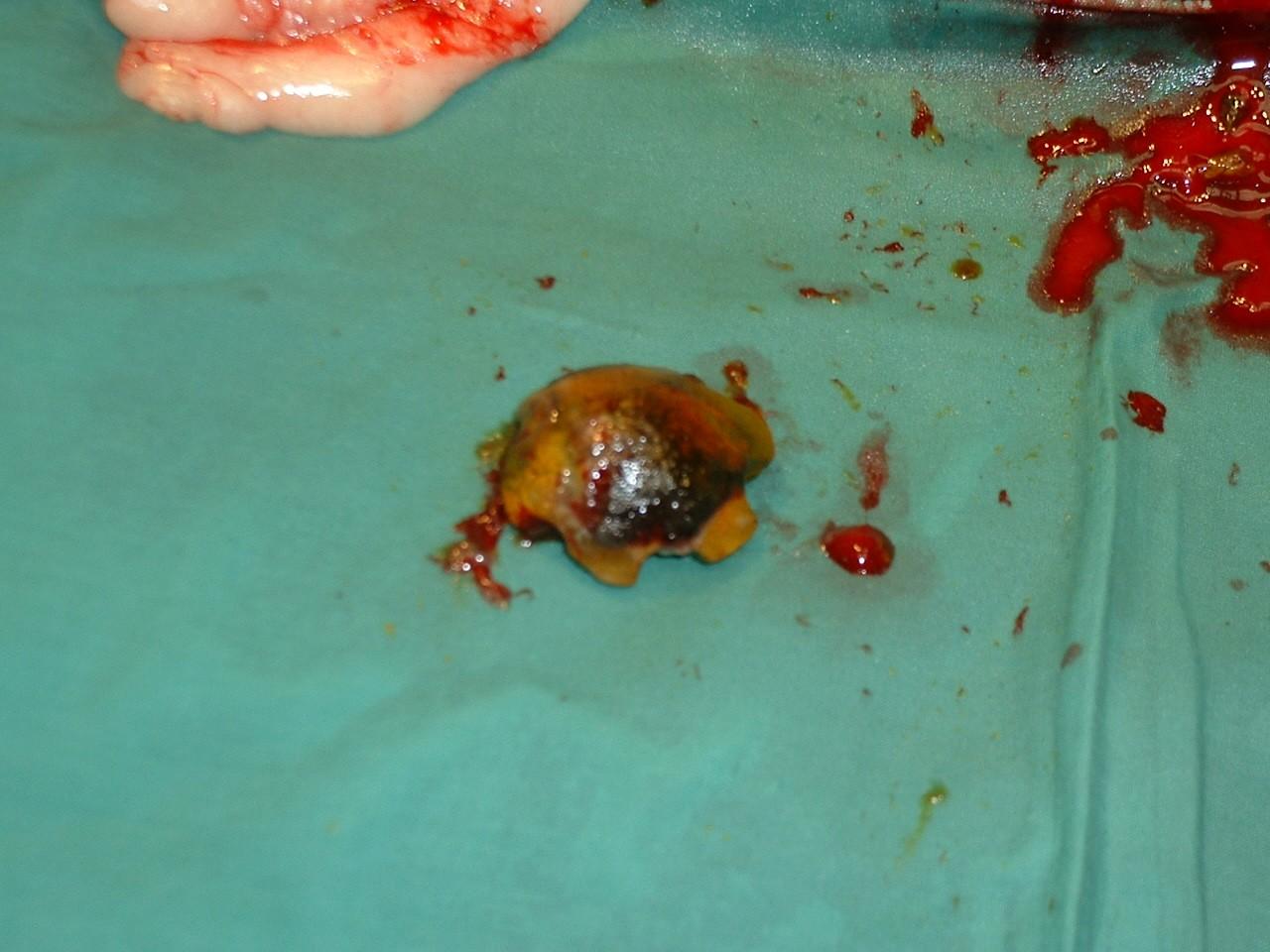
Exploratory laparotomy was performed based on the sonogram findings suggestive for intestinal obstruction and emerging perforation of various portions of small intestine. The surgeon found a 3 cm tubular rubber duodenal foreign body and multiple regions of localized peritonitis in portions of small intestine on either side of the foreign body obstruction. Histopath: mild/moderate lymphoplasmacytic and eosinophilic enteritis. Concurrent minor lymphoplasmacytic gastritis was also present with evidence of Helicobacter infection.
Video
Patient Information
Patient Name :
Lacey C
Gender :
Female, Spayed
Species :
Canine
Type of Imaging : Ultrasound
Status :
Complete
Liz Wuz Here :
Yes
Code :
04_00066
Clinical Signs
- Diarrhea
- Lethargy
- Vomiting
Exam Finding
- Abdominal Pain
- Dehydration
Images
Blood Chemistry
- Albumin, Low
CBC
- Left Shift
- WBC, High
Clinical Signs
- Diarrhea
- Lethargy
- Vomiting