This 9-year-old MN DSH presented for weight loss and postprandial vomiting. The physical exam revealed a large, palpable cranial abdominal mass. Blood analysis revealed moderately elevated SAP, moderately elevated ALT, and moderately elevated bilirubin levels. CBC was normal.
This 9-year-old MN DSH presented for weight loss and postprandial vomiting. The physical exam revealed a large, palpable cranial abdominal mass. Blood analysis revealed moderately elevated SAP, moderately elevated ALT, and moderately elevated bilirubin levels. CBC was normal.
Case Study
Intestinal Lymphoma – High Grade in a 9 year old MN DSH cat
Sonographic Differential Diagnosis
Small intestinal wall mass with adjacent segment of bowel suggestive of inflammatory bowel disease. Hence, primary consideration is given to lymphosarcoma. Similar round cell tumors are less likely, adenocarcinoma, leiomyoma or leiomyosarcoma cannot be definitively ruled out.
Image Interpretation
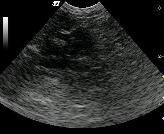
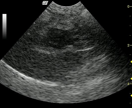
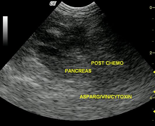
The first image demonstrates a hypoechoic, irregularly marginated mass asymmetrically arising from a loop of small bowel. There is also an echogenic linear tract within the described intestinal wall mass representing ultrasound guided tissue sampling with a 16 gauge Tru-cutВ® needle. Within the central portion of the mass, complete loss of bowel wall layering is noted while peripherally, the mass is continuous with a moderately to severely thickened muscularis layer suggesting these changes may represent malignant transformation of chronic inflammatory bowel disease to lymphosarcoma. Image 2: A continuous serosal margin suggests the mass is well contained and the adjacent soft tissue structures, most notably the pancreas are likely uninvolved. Image 3: partial reduction in the hypoechoic mass was noted post chemotherapy consistent with partial remission.
DX
High grade intestinal lymphoma
Outcome
The patient thrived for 7 months on a chemotherapy protocol (vincristine, asparaginase, Cytoxan, Adriamycin, prednisone) after which clinical decline led to euthanasia 8 months post diagnosis after presenting with a thoracic mass and pleural effusion.
Clinical Differential Diagnosis
GI pathology – Neoplasia, gastroenteritis; pancreatic pathology – pancreatitis,
Sampling
16 ga US guided biopsy of the mass revealed high grade lymphoma without evidence of originating tissue.
Video
Patient Information
Patient Name :
Chester M
Gender :
Male, Neutered
Species :
Feline
Type of Imaging : Ultrasound
Status :
Complete
Liz Wuz Here :
Yes
Code :
04_00051
Clinical Signs
- Vomiting
- Weight loss
Exam Finding
- Palpable mass
Images
Blood Chemistry
- Alkaline Phosphatase (SAP), High
- ALT (SGPT), High
- Total Bilirubin, High
Clinical Signs
- Vomiting
- Weight loss