This 3-year-old MN DSH was presented for weight loss. The physical exam revealed hypothermia and palpably enlarged kidneys. A CBC and blood chemistry profile demonstrated severe azotemia that decreased after fluid therapy. Moderate anemia was also present.
This 3-year-old MN DSH was presented for weight loss. The physical exam revealed hypothermia and palpably enlarged kidneys. A CBC and blood chemistry profile demonstrated severe azotemia that decreased after fluid therapy. Moderate anemia was also present.
Case Study
Intestinal and renal lymphoma in a 3 year old MN DSH cat
Sonographic Differential Diagnosis
Intestinal and renal lymphoma, carcinoma, mast cell disease, ulcerative inflammatory disease.
Image Interpretation
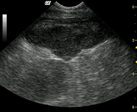
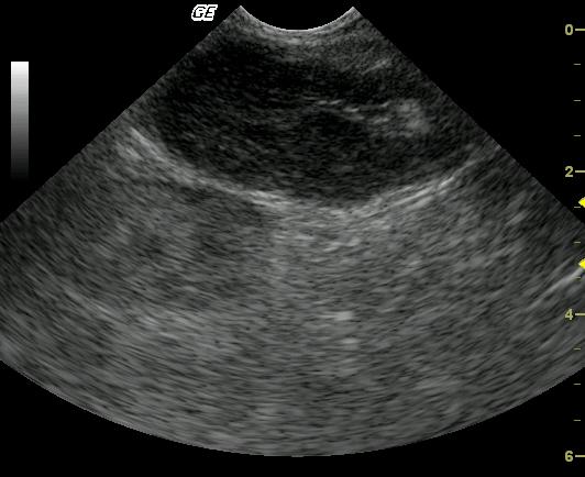
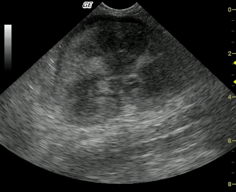
This image of a segment of bowel shows a markedly, asymmetrically thick wall with loss of layering and an undulating, although intact, serosal margin. Image 2 shows a moderate reduction of bowel wall thickness with subtle, peripheral return of normal wall layering. The kidney demonstrates a hypoechoic subcapsular ring with discrete loss of corticomedullary detail and mixed echogenic cortical changes.
DX
Intestinal and renal lymphoma
Outcome
BUN and creatinine were restored to normal limits, as was the hematocrit, under a CHOP protocol. The patient was doing well 3 months post diagnosis with a 2-pound weight gain and return to a strong clinical state.
Comments
This case presented for weight loss and enlarged kidneys on palpation. The intestinal segment was found to be abnormal as well and supported infiltrative disease. In the feline patient, the top three differentials included lymphoma, mast cell disease and adenocarcinoma. The renomegaly was suspected to be related and likely due to lymphosarcoma.
Clinical Differential Diagnosis
Bilateral renomegaly – Neoplasia, FIP, pyelonephritis, hydronephrosis, polycystic kidney disease, perirenal pseudocysts.
Sampling
22 g U/S guided FNA was performed of the hypoechoic region of mural detail loss in the intestinal mass (1 o`clock position video 1) and of the renal cortex. Cytology was consistent with lymphoma.
Video
Patient Information
Patient Name :
Stevie H
Gender :
Male, Neutered
Species :
Feline
Type of Imaging : Ultrasound
Status :
Complete
Liz Wuz Here :
Yes
Code :
04_00019
Clinical Signs
- Weight loss
Exam Finding
- Hypothermia
- Large Kidneys
Images
Blood Chemistry
- BUN high
- Creatinine, High
- GGT High
CBC
- RBC, Low
Clinical Signs
- Weight loss