1 year old DSH present for three day history of vomiting and not eating
– owner also thought that the patient may have been coughing a so rDVM thought maybe the cat may having been coughing leading to vomiting – history not clear
– bloodwork including FIV/FeLeuk Negative T-39.4C
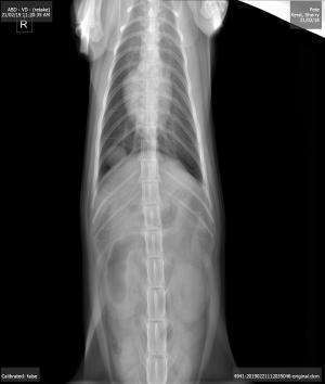
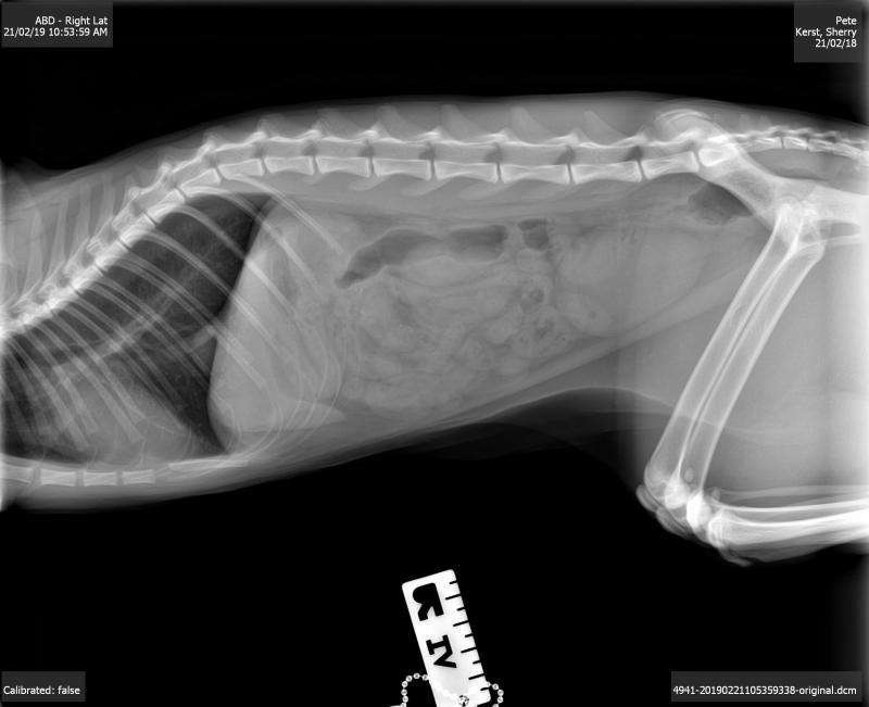
– x-rays show an approx. 1 cm nodule in the right caudal thorax
– the nodule could not be located on thoracic scan between the ribs even with different positioning and with collapsing the rib cage (the cat was heavily sedated)- only normal lung surface seen
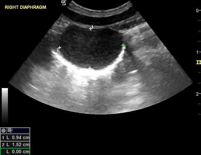
– the nodule could be seen however with a sub-xipohoid approach angling the probe cranially under the rib cage and to the right scanning through the liver and the diaphragm; colour flow not appreciated
– the aorta, cvc and esopahgeal inlet on the left side were all identified and not associated with the mass
– rest of abdominal scan normal except there was some food in the stomach so we suspect the patient has been eating a bit
So what is this? Sampling not possible as lung sliding in and out cranially and liver sliding caudally over the region so risk of laceration. To me it appears to be attached to the thoracic side of the diaphragm and not in the lung. I am not even sure if this is incidental at this time.


Comments
Looks there is fluid and
Looks there is fluid and hyperechoic material within the mass and with the pyrexia, anorexia, and coughing would considered an intra-thoracic abscess or granuloma
abscess or granuloma as remo
abscess or granuloma as remo says… the rad looks like its lung but the diaphragm seems to go around it which would mean liver but may just be the angle. Needs a needle for sure.
This is definitely not in the
This is definitely not in the liver but could be on the abdominal side of the diaphragm – just hard to tell and there is just no way I can get to this with a needle with the position of the probe deep subcostal pointing up.
We are going to try antibiotics and rescan. CT would be nice!
Jacquie
Jackie
The first thing that I
Jackie
The first thing that I would try is to get ( I am sorry to say ) an image with a linear probe. You need to first evaluate the nature or appearance of the encapsulating wall. Is it thickened? Is it a single layer as would be expecteted with a benign cystic stucture? Is the tissue surrounding it along all of it circumference quiescent of angry? These are all important things to know in advance.
The other really important point is whether or not the contents of the structure are echogenic or not. I am not sure if what appears to be echos within it are true or not. This will help differentiate between a plain cyst or an early abscess. If it is an abscess I would also expect to see changes in the parenchyma of the lung where you are making contact. I don’t see this happening in these images.
Finally if you can image it this proximally then knowing your skills, you should be able to aspirate it as per Eric’s suggestions. I would go for it if it looks like an abscess. The immediate benefit is draining of a large bacterial population which in itself is a bonus.
Hi Bob Thanks for your
Hi Bob Thanks for your input! If you could just see how I had to manipulate the probe under this patients ribs, subcostally, you would see both A) how difficult it was be to get a linear probe here (I tried and this is a cat) and B) The almost impossible placement of a needle without risking cutting my probe head as I would have to blindly slide the point up under the ribs in a position where the probe was so deeply buried in the tissue and pointing almost to the ceiling. The probe head completely disappeared in the tissue just to see the lesion. Just no room for a needle!
This nodule could not be seen intercostsally or from a proximal position at all which would have been the ideal position to get it but only gas shadow from lungs seen- even with deep manipulation. I am not afraid of using a needle in the chest or elsewhere but this his one had me beat I am sad to say : (
I totally get it. Was the pet
I totally get it. Was the pet sedated or under GA for this exam? If so different positions may have offered you more options. I know that you would have tried everything. I wasn’t trying being judgemental in any way. It just seemed to me if it was at all possible that it would be important to know if the contents were anechoic or echogenic. Nice job getting the images that you did.
No worries Bob! The patient
No worries Bob! The patient was well “kitty-magic-ed” out.