– 11 year old MN Shih Tzu with a chronic history of inteminttent vomiting (bile, foam) and signs consistent with possible GER
– pet has been treated with sulcrate, omeprazole, metoclopramide but has never really gotten better. The owners said the patient did better when placed on a trial of baytril and clavamox
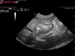
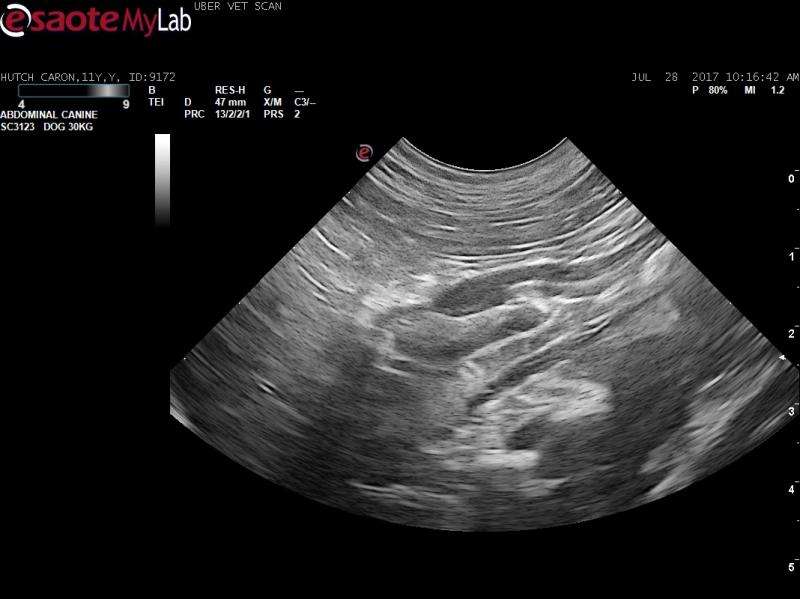
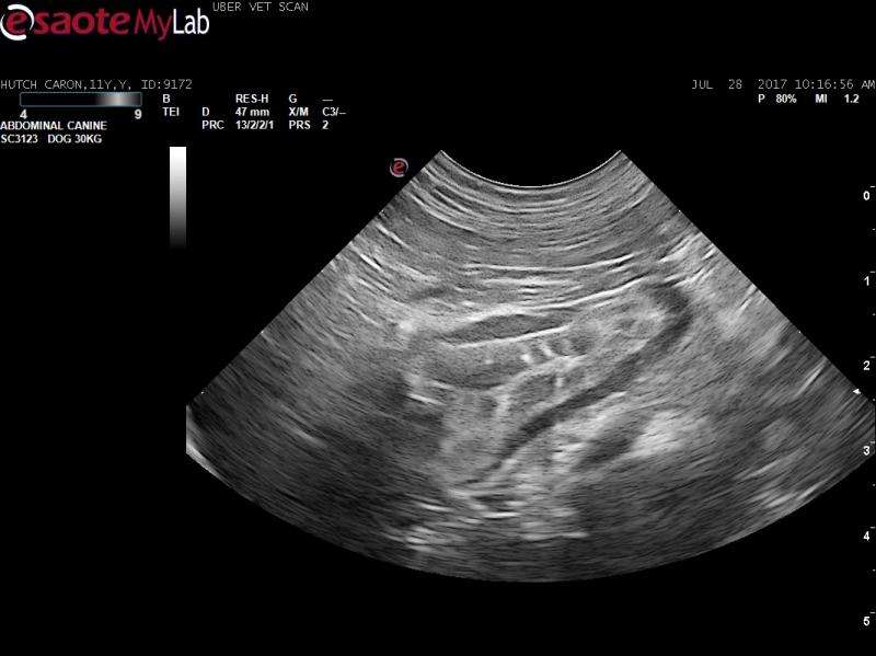
– abdominal ultrasound showed a gastric wall thickening at the pylroic outflow which appeared to affect primarily the mucosal layer; there are odd hyperechoic striations in the mucosa here
– endoscopy revealed a raised, hyperechoic mass-like lesion and biopsies were taken
– histopath indicated pyloric mucosal hypertrophy (possible pyloric stenosis) however a hyperplastic polyp can not be ruled out from the description I gave the pathologist
Any thoughts on the ultrasound images? The lesion appears more circumferential on ultrasound but seems more polyp-like on endoscopy. I suspect that this is the reason for the patient’s chronic vomiting. Not sure where to go from here?


Comments
Endosopic image and
Endosopic image and ultrasound more indicative of pyloric hypertrophty which is in line with the histopathology. There are surgical techniques to improve outflow pyloric outflow and would also do full thickness biopsy of the lesion.