I attempted to post yesterday but didn’t appear to publish. Sorry if this is a double post! I need some opinions on this case.
I attempted to post yesterday but didn’t appear to publish. Sorry if this is a double post! I need some opinions on this case.
- Signalment: 6 yo FS pit bull mix
- Acute onset vomiting, inability to keep food down
- No hx of medication admin or toxin ingestion
- Labwork shows anemia (mild), hypoproteinemia, increased liver values
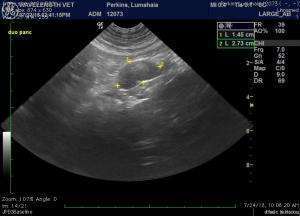
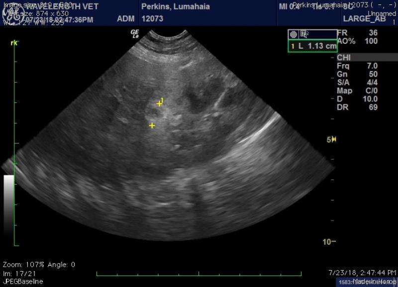
- U/s shows thickened wall with loss of layering in pyloric outflow, large gastric LN, multifocal “target” lesions through liver and hypoechoic lesions through spleen, irregular kidneys with small target lesions in cortex, and prominent iliac lymph nodes surrounded by inflammation.
- FNA of gastric LN, a target lesion in the liver and the pyloric wall lesion were reported as follows:
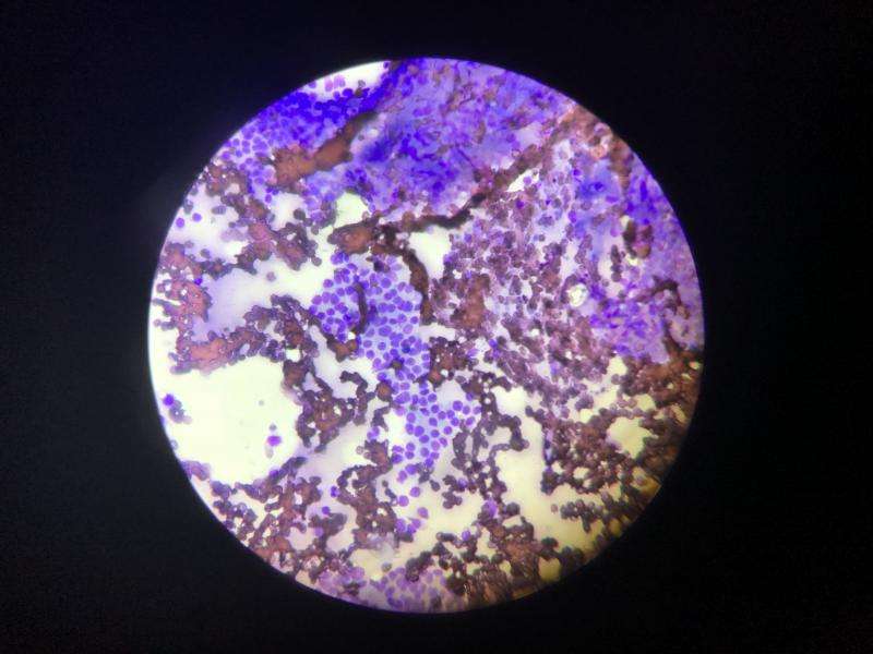
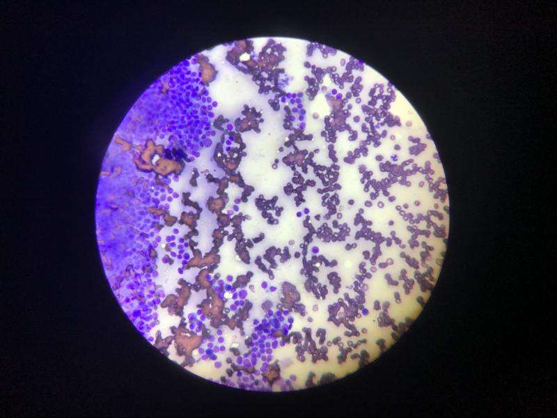
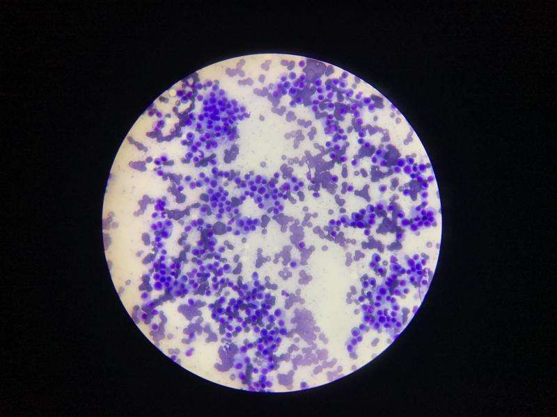
- – FNA of stomach (pic attached): cytology revealed limited evidence of mixed small lymphocytes and a pleomorphic plasma cell population (comments – poor cellular recovery per pathologist but the slides looked pretty cellular to me)
- – FNA of lymph node (hemorrhagic): cytology revealed hemorrhage with low numbers of a mixed suppurative, lymphocytic and pleomorphic plasma cell infiltrate. (comments – clinical significance is uncertain, possibly mildly inflamed and reactive lymph node)
- – FNA of liver lesion: significant numbers of a pleomorphic plasma cell population with few small lymphoid cells, suggestive for underlying plasma cell myeloma. (Comments – there are no hepatocytes observed on the slides).
Needless to say, the cytology read is frustrating to me. The liver lesion was overtly in the liver, so I don’t know why there weren’t hepatocytes – maybe I should’ve been more on the margin of the lesion? The stomach slide was very cellular but only in certain locations (First two cytology photos are stomach wall, third is liver lesion).
The dog has been euthanized due to intractable vomiting despite supportive care, but I would love to give these owners more of a definitive answer. I would welcome any thoughts!
Liz
Comments
Hepatocytes from liver FNA.
Hepatocytes from liver FNA. Always look for the nucleated cells when snapping pics or videos of cells. Also what magnification are you using? We take images at 10X, 40X, with no oil, and then 100X with oil immersion.
This may be helpful: https://sonopath.com/resources/spa-sonopath/telecytology-services/sdep-telecytology-samples-cell-collections-what-look-yo
Thanks Kelly – Yes, there are
Thanks Kelly – Yes, there are lots of nucleated cells in those slides. It’s 40x. I did not want to put oil on the slides as we were submitting the slides and oil makes Antech pathologists angry. 🙂 I was pushing my luck sending them stained slides as it was! Haha.
Oh my gosh really? The images
Oh my gosh really? The images are so nice with the oil immersion. 🙂
Not if you send to the lab
Not if you send to the lab with oil and it dries. It’s hard to read then – blurry under the scope. To clarify: This was an old-fashioned, send the slides to the lab cytology read, not telecytology. I can blow up on my phone and repost if that would be helpful.
Oh, I see that makes more
Oh, I see that makes more sense. 🙂
Kelly can you flag this for
Kelly can you flag this for one of the docs to take a look? Thanks! 🙂
Here’s a zoomed in pic of the
Here’s a zoomed in pic of the stomach cytology
Im not a cytologist but looks
Im not a cytologist but looks like round cell neoplasia to me and certainly the sonogram suggests this.
Maybe do a telecytology with our DR McGill?
Thanks Eric – Unfortunately I
Thanks Eric – Unfortunately I blew the client’s budget on a very unhelpful cytology read by Antech, and now the dog is no longer with us, so….Tried for a second opinion at Antech and found them still saying that the slide wasn’t very cellular. Sure looks adequately cellular to me!
Looks pretty cytological to
Looks pretty cytological to me….
Any cyto wizards in the
Any cyto wizards in the group?