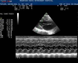
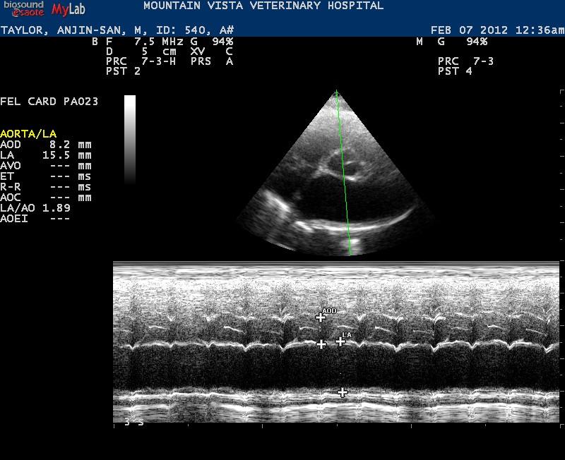
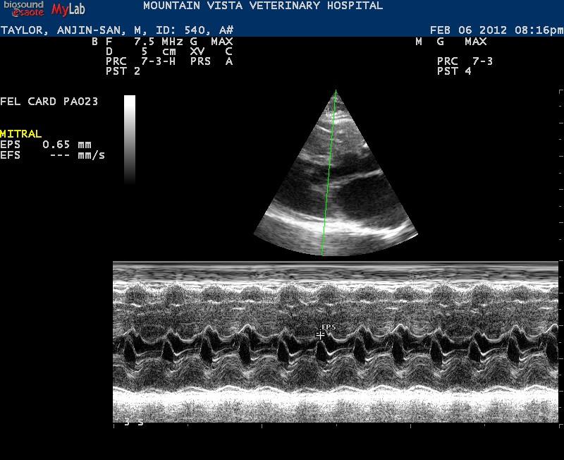
– 2yr old MN DSH presented for respiratory distress
– no heart murmur detected T=33C!
– chest rads show suspected pulmonary edema and enlarged heart
– treated with furosemide and oxygen support
– performed echoe once deemed stable (but was very tricky!)
– gave low dose butorphanol to help calm and did my best!
1. Any hints on performing meaningful echoes on these patients that are on the edge?
2. The clip of the LV shows a strange lesion in the IVS (looks like 2 anoechoic nodules) – is this a true lesion or just poor positioning on my part creating this?
3. What criteria do you use to rule out SAM? (I don’t think this pet has it – had trace MR on colour doppler, aortic velocity 0.89)
Our kitty on presentation lateral thorax
[videoembed id=6919]


Comments
Nice scan Jacquie, I dont see
Nice scan Jacquie, I dont see SAM here. Regarding sam see my and Peter’s case of the month form december 2011. HCM yes and assymetric hypertrophy, thickened mv. La/ao eyeballing about 2.0 and a shower curtain lung pattern at 11 o’clock on the clip indicated pulmonary edema but pte, pneumonitis, neoplasia can do the same but given the big la pulmonary edema is likely the scenario +/- lung clots especially if persistent. I dont get big into measurements on these guys early. The issue with respiratory distress echo is volume overload vs non. In this case there is volume overload and la/ao is excessive and contractility high with thick LV. HCM with lae and chf. Cage rest, 02, lasix, nitro if you like it, opioids +/- antithrombotics. Once they are out of failure and stable, if that occurs then reecho and put a name on it. They often change their parameters in CHF and when out of CHF so UCM or DCM (not this case) becomes HCM and vice versa. Semantics mean nothing if stressing a cat over the edge puts him in the freezer. Just put the probe on get the views pressing video clips (p1 on the logic e ) and get him in a cage and post process the images later. Torb him IM 30 minutes prior as this will calm him down and avoid excessive o2 consumption without affecting the heart function. The key to emergency US is just being quick and using technology to “get what you need”…. Mick Jagger echo:)
Re Q2: I think this is just a
Re Q2: I think this is just a form of hcm and hypertrophy with fibrosis or similar (hyperechoic muscular lesion). Let me see if Peter can chime in.
Re Q2: I think this is just a
Re Q2: I think this is just a form of hcm and hypertrophy with fibrosis or similar (hyperechoic muscular lesion). Let me see if Peter can chime in.
I totally agree with Eric.
I totally agree with Eric. Once you got an estimation of the LA size it´s not important to put a name on the cardiomyopathy until the patient has been stabilized. I usually use a lasix-CRI at 1 mg/kg/hr, O2 and – if necessary – a sedation (acepromacine/butorphanol). If there was markedly decreased systolic function seen on the overview echo I add dobutamine as a CRI.
Regardin you lesion: I think the hyperechoic structure is a false tendon crossing the LV and the lesion behind it is just a part of the LV , but I cannot tell it for sure anyway. I would repeat the echo once the patient has been stabilized.
Regarding SAM; I cannot see it on the material provided. You got a good machine and therefor should be able to see it in 2D views (reduce sector angle to increase frame rate). You can use CDI either (turbulent flow across the LVOT and mitral regurg originating from the same point). M-modes can also help. But basically SAM causes a heart murmu.
Regarding your LV measurements: Be sure not to include partes of the papillary muscles or the RV into your IVS or PW measurements….
Hope I could help!
Best Regards from Austria!
Peter
Thanks Peter
I was not 100%
Thanks Peter
I was not 100% confident with my LV wall measurements on the m-mode (often find them difficult even in normal cats because of the papillary muscles). Thanks for the tips. I also do 2-D measurements to get another measurement to compare if I can. I know HCM can be easily over diagnosed.
On a separate note, I see on some of your images on Sonopath that you use a MyLab machine. What is your opinion on the PA240 probe? It looks like it produces some very nice images. I currently have a PA023 and PA121.
Jacquie
Thanks Peter
I was not 100%
Thanks Peter
I was not 100% confident with my LV wall measurements on the m-mode (often find them difficult even in normal cats because of the papillary muscles). Thanks for the tips. I also do 2-D measurements to get another measurement to compare if I can. I know HCM can be easily over diagnosed.
On a separate note, I see on some of your images on Sonopath that you use a MyLab machine. What is your opinion on the PA240 probe? It looks like it produces some very nice images. I currently have a PA023 and PA121.
Jacquie
Hi Jacquie!
Yes, 2D
Hi Jacquie!
Yes, 2D measurements are good but be aware of the frame rate (should be quite high). Regarding the probe: I use the 023 and 121 as well as the 240 for cardiology. The advantage of the 240 is the low frequency which makes it very useful in large breed dogs and doppler measurements (high velocities, CDI). I like it very much. But the 230 is also a good probe….
Using the 121 for large breeds often leads to underestimation of regurgitation based on CDI. And the maximal velocity you can measure is limited.
In cats I sometimes use the microconvex probe (I know a cardiologist should not do that, for what reason ever…). I makes brilliant 2D images. It is not a good probe for measurements because the frame rate is not that high but it makes you see myocardial abnormalities very clearly.
Best Regards!
Peter
Hi Jacquie!
Yes, 2D
Hi Jacquie!
Yes, 2D measurements are good but be aware of the frame rate (should be quite high). Regarding the probe: I use the 023 and 121 as well as the 240 for cardiology. The advantage of the 240 is the low frequency which makes it very useful in large breed dogs and doppler measurements (high velocities, CDI). I like it very much. But the 230 is also a good probe….
Using the 121 for large breeds often leads to underestimation of regurgitation based on CDI. And the maximal velocity you can measure is limited.
In cats I sometimes use the microconvex probe (I know a cardiologist should not do that, for what reason ever…). I makes brilliant 2D images. It is not a good probe for measurements because the frame rate is not that high but it makes you see myocardial abnormalities very clearly.
Best Regards!
Peter
Thanks Peter
All very
Thanks Peter
All very helpful information. I will definitely be paying more attention to frame rates now!
I have been having some colour flow problems with the 121 (not really happy with strength of the signal in some pets despite adjusting PRF, frequency, depth, scan width etc.) So I have been considering investing in the PA240 probe but it is nice to finally speak to someone who is actually using it! Unfortunately our MyLab dealer in Canada has not been overly helpful with my questions.
Jacquie
Thanks Peter
All very
Thanks Peter
All very helpful information. I will definitely be paying more attention to frame rates now!
I have been having some colour flow problems with the 121 (not really happy with strength of the signal in some pets despite adjusting PRF, frequency, depth, scan width etc.) So I have been considering investing in the PA240 probe but it is nice to finally speak to someone who is actually using it! Unfortunately our MyLab dealer in Canada has not been overly helpful with my questions.
Jacquie
Hi Peter (and
Hi Peter (and Jacquie),
Peter, when you use the 023 probe, does it give good doppler and 2-D images on cats and small dogs? I also have a Mylab. Currently I use the CA123 for 2-D on these smaller patients, and the PA121 to get the doppler readings. It’s frustrating though, because the PA121 gives horrible 2-D images in some of them.
It would be nice to use one probe.
Haven’t tried the PA240. I find the PA121 has given good images on all the larger dogs. Interesting though your comment about it’s underestimation of regurgitation in CDI. I must remember that.
Karen
Hi!
The P023 gives
Hi!
The P023 gives brilliant images on cats and small dogs, also CDI and Spectral Doppler (CW does not work on the MyLab70 however, but is does on My Lab 30. 40 and 50). The 121 is good for medium sized dogs, also with regard to Spectral and CDI. Due to physical reasons it´s always better to use a lower frequency for CDI than for 2D, that´s why I use teh 240 also for CDI in medium sized dogs. You can achieve perfect 2D images in large breed dogs using the 240. If you want to use high velocities in dogs (e.g. SAS, PS), it´s better to use the 240 probe to avoid aliasing.
The micro-convex probe is useful to evaluate myocardial abnormalities in cats but it´s less useful for Doppler.
I would recommend contacting your local Esaote representative. Perhaps he can provide you with the probes for some days so that you can check out if you like them.
Best Regards
Peter
Hi!
The P023 gives
Hi!
The P023 gives brilliant images on cats and small dogs, also CDI and Spectral Doppler (CW does not work on the MyLab70 however, but is does on My Lab 30. 40 and 50). The 121 is good for medium sized dogs, also with regard to Spectral and CDI. Due to physical reasons it´s always better to use a lower frequency for CDI than for 2D, that´s why I use teh 240 also for CDI in medium sized dogs. You can achieve perfect 2D images in large breed dogs using the 240. If you want to use high velocities in dogs (e.g. SAS, PS), it´s better to use the 240 probe to avoid aliasing.
The micro-convex probe is useful to evaluate myocardial abnormalities in cats but it´s less useful for Doppler.
I would recommend contacting your local Esaote representative. Perhaps he can provide you with the probes for some days so that you can check out if you like them.
Best Regards
Peter