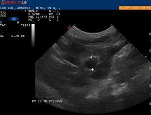
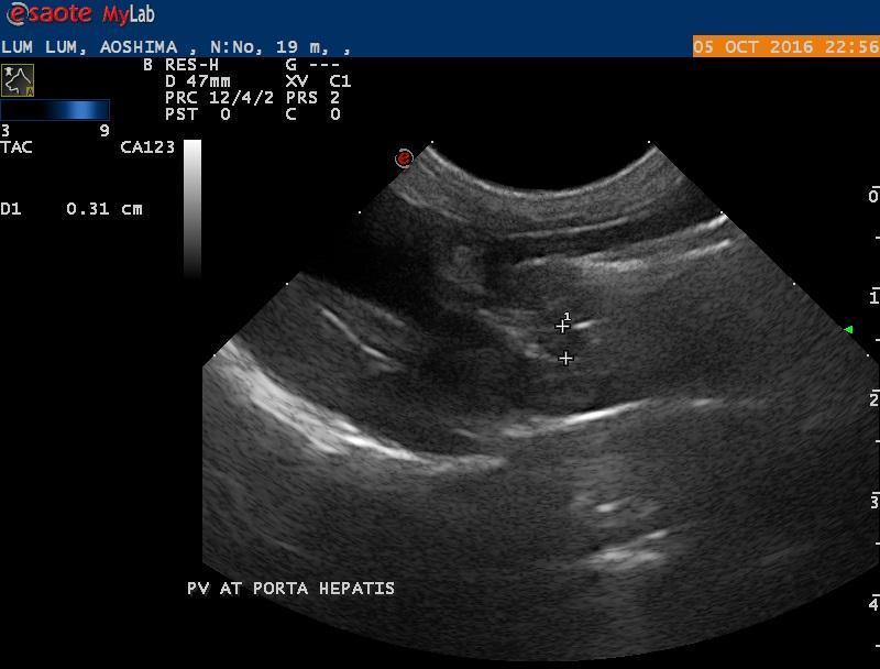
– 19 months old, Female entire Lhasa Apso. Presented for scan to check that she was not neutered. Incidental finding: severe microhepatica, change in diameter of portal vein from caudal to pylorus to porta hepatis levels. Kidneys appear WNL in size, with hyperechoic cortical band and minor calculi/mineralized focci. Rest WNL. Biochemistry WNL.
– 19 months old, Female entire Lhasa Apso. Presented for scan to check that she was not neutered. Incidental finding: severe microhepatica, change in diameter of portal vein from caudal to pylorus to porta hepatis levels. Kidneys appear WNL in size, with hyperechoic cortical band and minor calculi/mineralized focci. Rest WNL. Biochemistry WNL.
– A gastroazygus shunting vessel is presumptively seen. Adviced to check postpandrial bile acids for reassurance. Test was performed 2 hours after a normal meal (non-fatty). Bile acids were within normal limits. My colleague asks how sure am I that there is a shunt…I’m pretty sure but I would need a second opinion as a CT scan might be a bit too expensive for confirmation (patient is totally asymptomatic…of course).
-Questions:
1- Do you think there is a shunt (apologies for the lack of transverse view but the clip I have is not very diagnostic, so I omited it).
2- In these scenarios, what other tests appart from CT can we do for further diagnosis? Definiitve diagnosis is only CT?
3- What is the risk of surgery (spay, or simple proceedures) when the patient is suspected of shunt but further testing is not possible due to financial reasons
 ?
?
4- How does a shunt affect chemotherapy?(this is a “by the way” question, as I have 2 cases with diagnosed lymphoma and shunts).
Thanks for any input.
Comments
Looks like more of a
Looks like more of a splenoazygos because gastroazygos has a looping pattern near the pylorus wheras splenic comes off the portal vein and heads dorsally like this one. Azygos because the cvc is normal size so the only other vessel to dump into is the azygos. Confirm with the double aorta sign cranial to the diaphragm.
See the attached shunt diagrams for the clinical approach book that we may finish sometime before the next century:)
And here’s a shunt search in the archive as there are multiple expamples of all types on US and CT for your research…. Man I love our archive:)
Thanks Eric. You shared those
Thanks Eric. You shared those patterns before and I find them very helpful. In some clips I thought there was a loop, not very clearly. But yes, may be splenoazygus would fit more;But what I wanted to confirm was the shubt itself. Thank you.
Any ideas on the other questions?