– 4 year OLD FS DSH presented for dyspnea and pleural effusion on x-ray
– had a history of cough a few weeks prior to presentation and history of chronic diarrhea with blood present
– 140ml serosaguineous fluid removed from chest; in house cytology showed small clusters of big purple cells with a large nuclei (I am no cytologist but malignant looking); official cytology pending
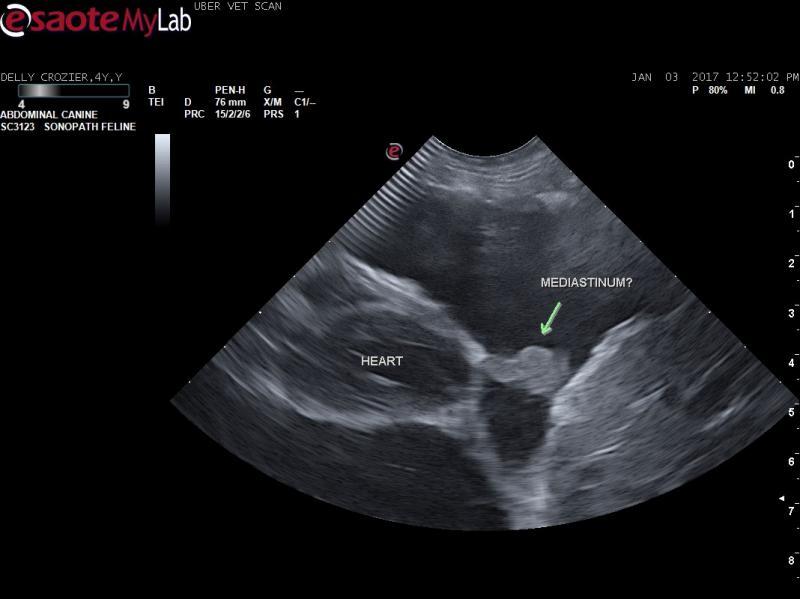
– a mediastinal mass was not seen on chest ultrasound but lung consolidation and possible lung mass; I am wondering if I am seeing thickened mediastinal tissue however?) (see images) LA/Ao wnl
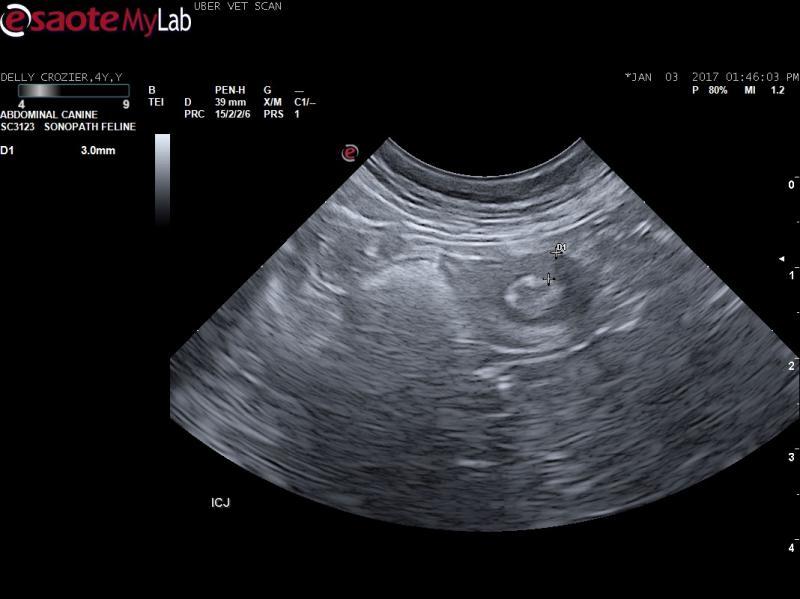
– thickened muscularis layer in ileum at ICJ and the liver had distended hepatic veins on abdominal ultrasound; moderately enlarged jejunal LN
So, with no mediastinal LN enlargement, I am placing lymphoma lower on the list – could this be lung carcinoma, FIP, mesothelioma, lung torsion?
The ileal lesion: lymphoma, localized FIP, IBD?


Comments
To start we know the pleural
To start we know the pleural effusion is non cardiogenic given the small LA and volume contraction. Your last video shows lung consolidation with intraparenchymal air interface which means lung origin to the mass as opposed to LN. Cells in clusters with this type of presentation I would be concerned for lung carcinoma/thoracic carcinomatosis.
You can telecytology your samples uploading to sonopath as well if you wish. For the protocol contact info@sonopath.com. We do all our cytology this way in the sonopath mobile practices.
http://spa.sonopath.com/
Thank -you EL. Do you think
Thank -you EL. Do you think the ileal lesion is a separate entity?
FYI – cytology from thoracic fluid confirmed carcinoma
i think the ileocecal is a
i think the ileocecal is a separate issue and likely not neoplasia