Rusty: 11 Year Old DSH
Presents for ADR and anorexia x3 weeks.
PE: 3/6 systolic murmur and fullness to abdoment
LABS: WNL except 21,500 WBC#
Abd. U/S: Moderate/Lg Volume ascites (analysis suggests mod. transudate). SI thickened w/ muscularis layer approaching thickness of mucosa and corrugation of SI present.
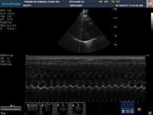
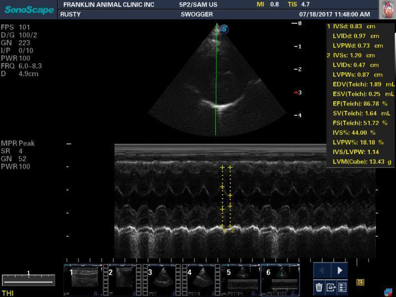
Card. U/S: LV walls thickened past 6mm in diastole but function appears normal w/ no SAM evident & only mild/mod increase in LA size.
Impression: ascites not due to CHF or RCHF and likely due to SI disease. Is this a correct reading, especially of the cardiac side of things?
Thanks,


Comments
There is also mucosal
There is also mucosal stippling of the intestine, which is indicative of lymphangectasis and severe IBD. However would then expect hypoalbuminemia with secondary ascites if low enough. The LV thickening >6 mm is indicative of myocardial hypertophy and together with enlarged La and murmur, most likley dealing with HCM/systemic hypertension/hyperthroidism. I assume that the latter 2 have been excluded.
What did the fluid cytology show?
Thanks for the reply,
The
Thanks for the reply,
The cytology had a TP of 2.8 and was mostly RBC’s, some reactive mesothelial cells and small lymphocytes.
Thyroid levels were WNL and I know my associate was checking BP, but I’ve not heard what that was yet.
Regarding the cardiac U/S…even with the LV wall thicknesses being increased, it doesn’t seem like the LA nor the function of the heart is that severly effected (although I may be underestimating the LA size…I admit cats are hard for me to evaluate). I would think it would take much more significant pathology to create a large volume ascites???
Thanks again for the help.
Sam
if you want to know if
if you want to know if ascites is from cardiac just look at the hepatic veins.. if they are not jumping out and are 1:1 with the portal veins and the CVC is 1:1 or less with the aorta then the thorax including the heart is not a player. Ascites from passive congestion must have a biug cvc and big hepatic veins. If those arent present then stay south of the diaphragm for the answer. I would be concerned for lymphomatosis/cacinomatosis or causes of lymphatic obstruction here that often dont exfoliate Images are a bit dark. Check for target liver lesions, spleen > 1 cm, ill defined pancreatic nodules and LNs.
Here are some passive congestion patterns with dilated HV and CVC with secondary ascites. Just put “passive congestion” as key words in the clinical search
http://sonopath.com/members/case-studies/search?text=passive+congestion&species=All
Thanks for the thoughts and
Thanks for the thoughts and advice
Sam
You bet Sam! I also recorded
You bet Sam! I also recorded “working through ascites” downloadable lecture currently there in products. This explains how to tell where the fluid is coming from with a few simple parameters and sonographic aspects of the organ systems. Lots of quick bullet tips like the hepatic vein discussion above.
https://sonopath.com/products/downloadable