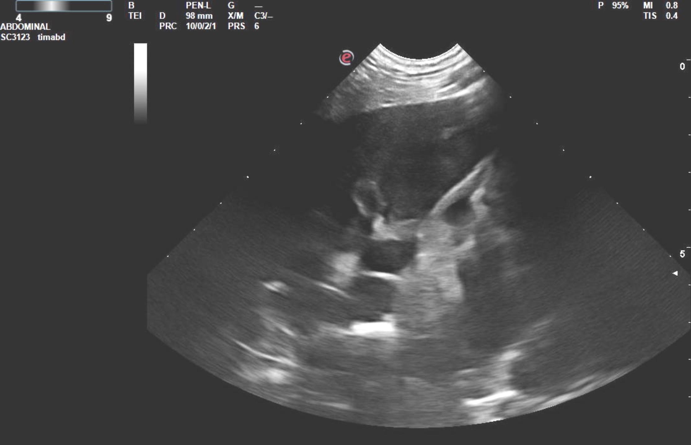
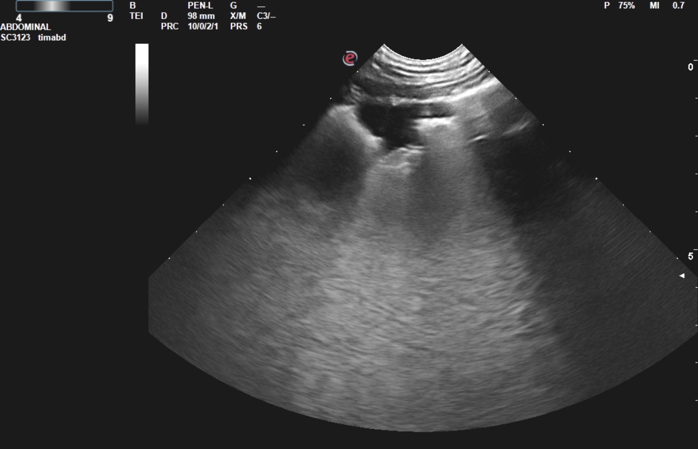
US/RAD/CYT – Lung fungal pneumonia confirmed as blastomycosis on FNA in a 5year old MN Weimaraner

Case Study

US/RAD/CYT – Lung fungal pneumonia confirmed as blastomycosis on FNA in a 5year old MN Weimaraner

This 5 year old MN Weimeraner has a history of 10 days of coughing, pyrexia (105.5), poorly responsive to antibiotics. No crackles on auscultation.

This 5 year old MN Weimeraner has a history of 10 days of coughing, pyrexia (105.5), poorly responsive to antibiotics. No crackles on auscultation.

Sonographic Differential Diagnosis

Differentials include sarcoma, pneumonia, carcinoma, possibly lung necrosis.

Image Interpretation

On ultrasound, focal lung consolidation was noted in this patient consistent with primary lung mass. 

Thoracic Rads: 


 There is moderate left mediastinal shift. There is increased opacity associated with the cranial subsegment of the left cranial lung, noted as increased opacity dorsal to the heart base on the lateral view, including a sharp margination of the caudal dorsal margin of the lung lobe (lobar sign) as well as small air bronchograms more cranially and ventrally. There may be some involvement of the caudal subsegment of the left cranial lung creating increased opacity superimposed over the mid cranial cardiac silhouette on the lateral view. The cardiac silhouette is appropriate for volume and margination. No pleural space disease is noted. No cranial mediastinal pathologies identified. Dorsal to the second sternebra there is a broad-based, convex soft tissue opacity that could indicate mild sternal lymphadenopathy 

Outcome

normal echo.
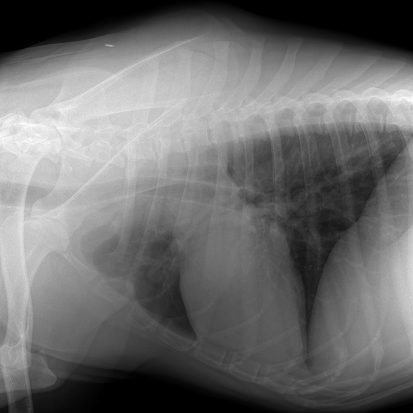
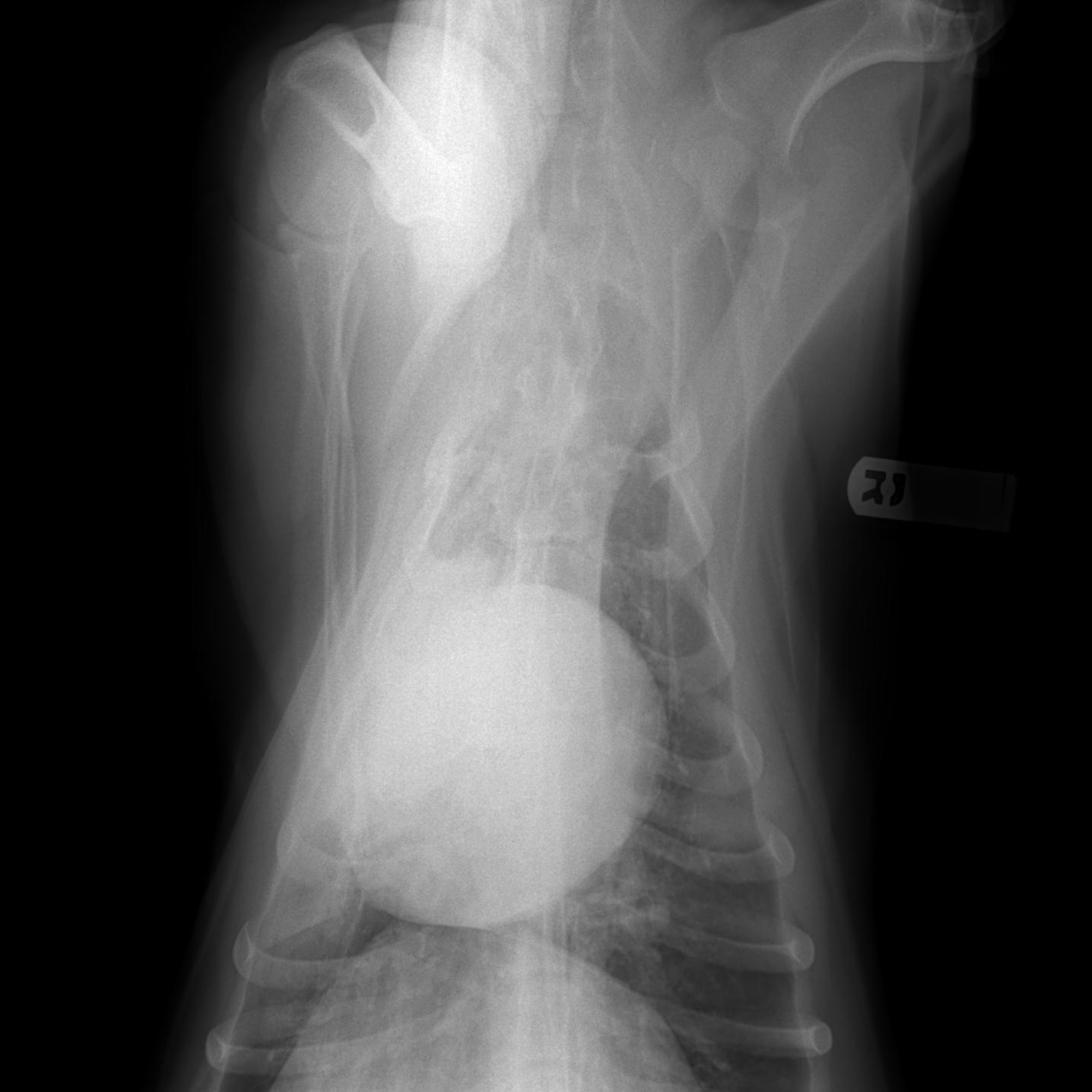
Thoracic rads: 1) Increased opacity, including air bronchograms, associated with the left cranial lung suggests pneumonia. Atelectasis may be playing a component role in this appearance, based on the left mediastinal shift noted on the VD view. Given the poor response to antibiotics, fungal pneumonia or an underlying inhaled foreign body could be considered. Neoplasia is less likely as typically, expansion of the affected lung lobe occurs, not the case with this patient.
2) Suspect mild sternal lymphadenopathy. Often, sternal lymphadenopathy is an indicator of abdominal pathology as these lymph nodes draining the peritoneal cavity.

Sampling

FNA of the lung consolidation – yeast infection consistent with Blastomycosis

Patient Information

Patient Name : Watson Rohen/Bayshore
Gender : Male, Neutered
Species : Canine
Type of Imaging : Ultrasound
Status : Complete
Liz Wuz Here : Yes

Clinical Signs

  • Coughing
  • Fever

Images

blasto_watson_latblasto_watson_vdwatson_lung_fungal_penumoniawatson3watson_40x_imagewatson_100x

Clinical Signs

  • Coughing
  • Fever
Skip to content