Thickened ventral bladder wall and apex, R/O chronic cystitis, TCC , or round cell in a 9 year old FS Pug mix

Case Study

Thickened ventral bladder wall and apex, R/O chronic cystitis, TCC , or round cell in a 9 year old FS Pug mix

A 9 year old FS Pug Mix was presented for evaluation of hematuria over a 2 month period that appeared to respond to Simplicef. Prior urinalysis had shown a specific gravity of 1.012 and prior blood work had shown for mild hyperglobulinemia as the only abnormality.

Physical examination was normal.

A 9 year old FS Pug Mix was presented for evaluation of hematuria over a 2 month period that appeared to respond to Simplicef. Prior urinalysis had shown a specific gravity of 1.012 and prior blood work had shown for mild hyperglobulinemia as the only abnormality.

Physical examination was normal.

Sonographic Differential Diagnosis

Thickened ventral bladder wall with loss of detail and thickened apex of the bladder.

Image Interpretation

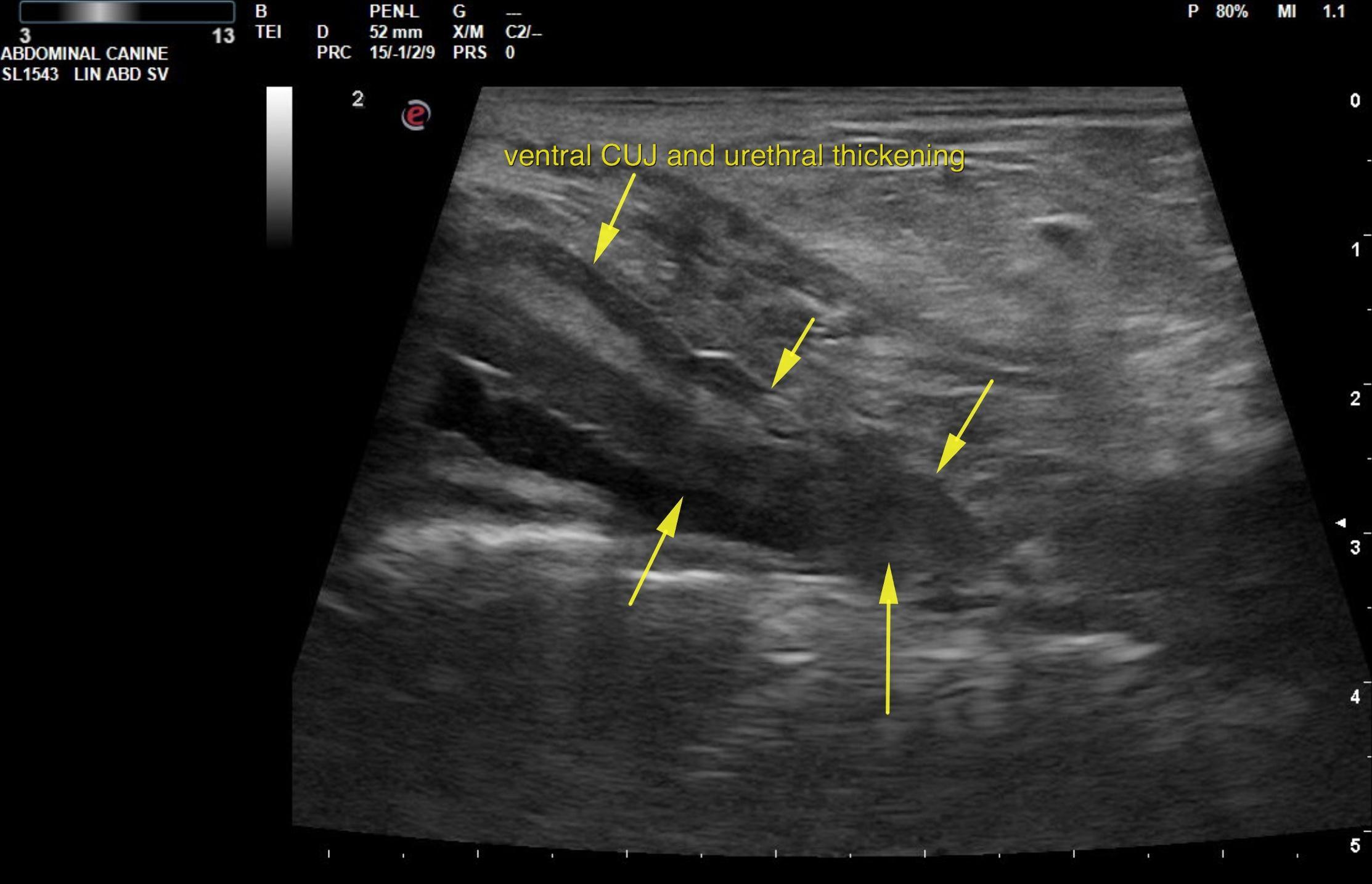
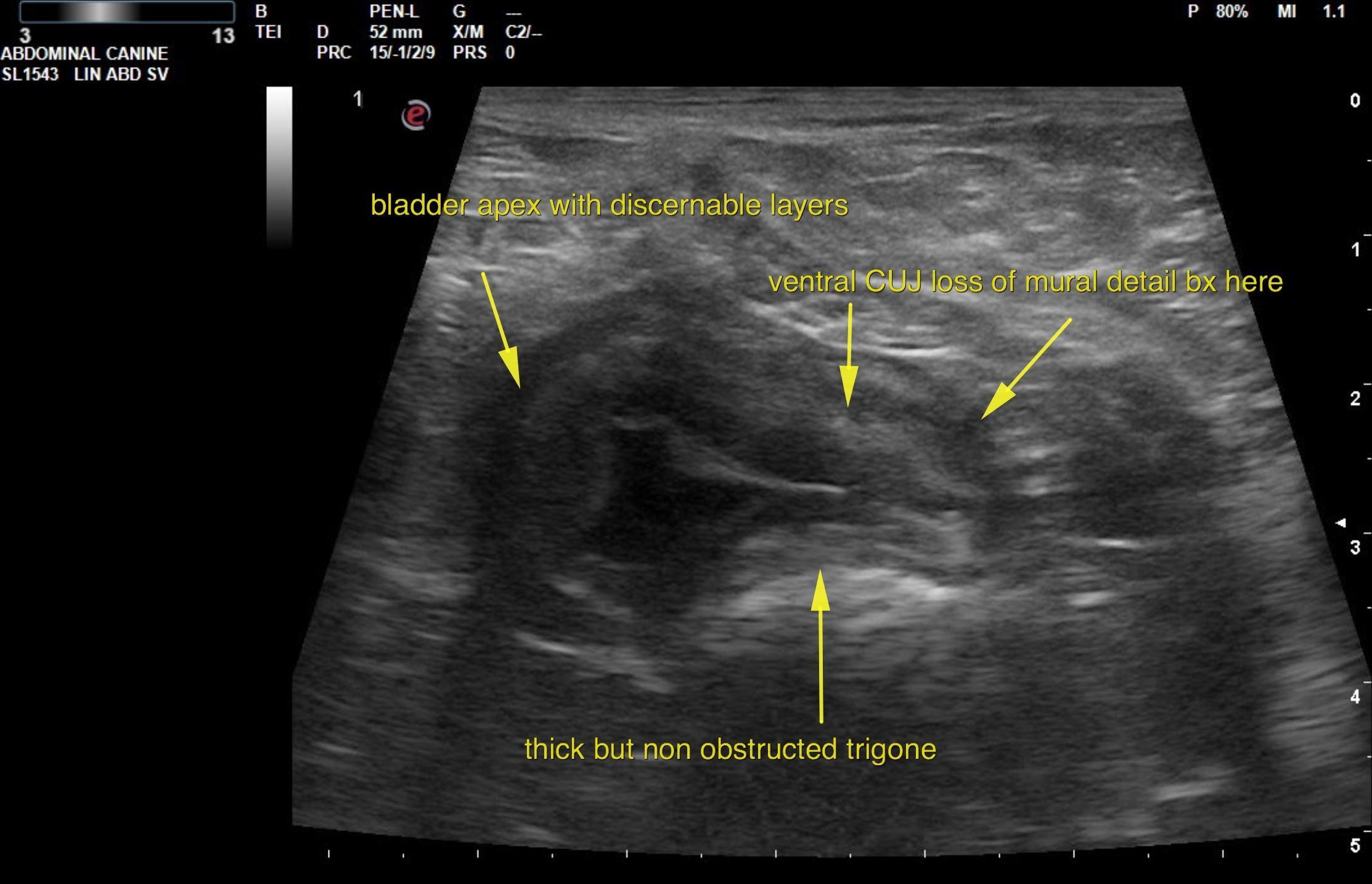
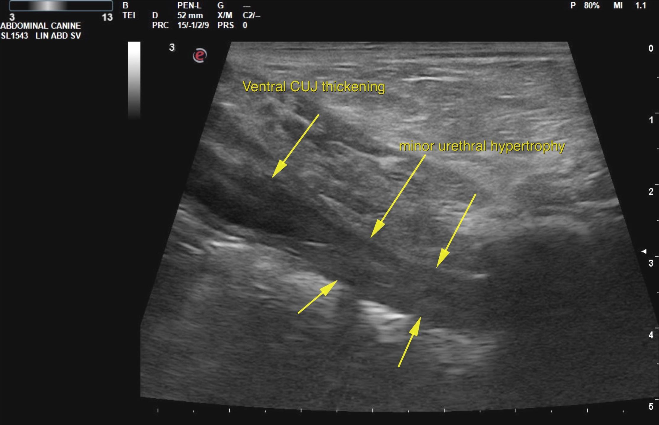
The urinary bladder presented thickened ventral wall that entered into the cystourethral junction and proximal urethra. Loss of mural detail was noted. Irregular serosal contour was noted. The apex of the bladder was also thickened and a minimal amount of urine was present. Therefore, this is affecting the degree of thickness in the bladder wall. However, sectorial hypertrophy was noted throughout the ventral apical wall and mildly in the dorsal wall. The ureters were not obstructed. The apex of the bladder had discernable layers with serosa, muscularis and mucosa all of which were thickened and hypertrophied. However, the caudal ventral aspect of the bladder is most concerning, particularly in the cystourethral junction region where irregular loss of mural detail was noted. 

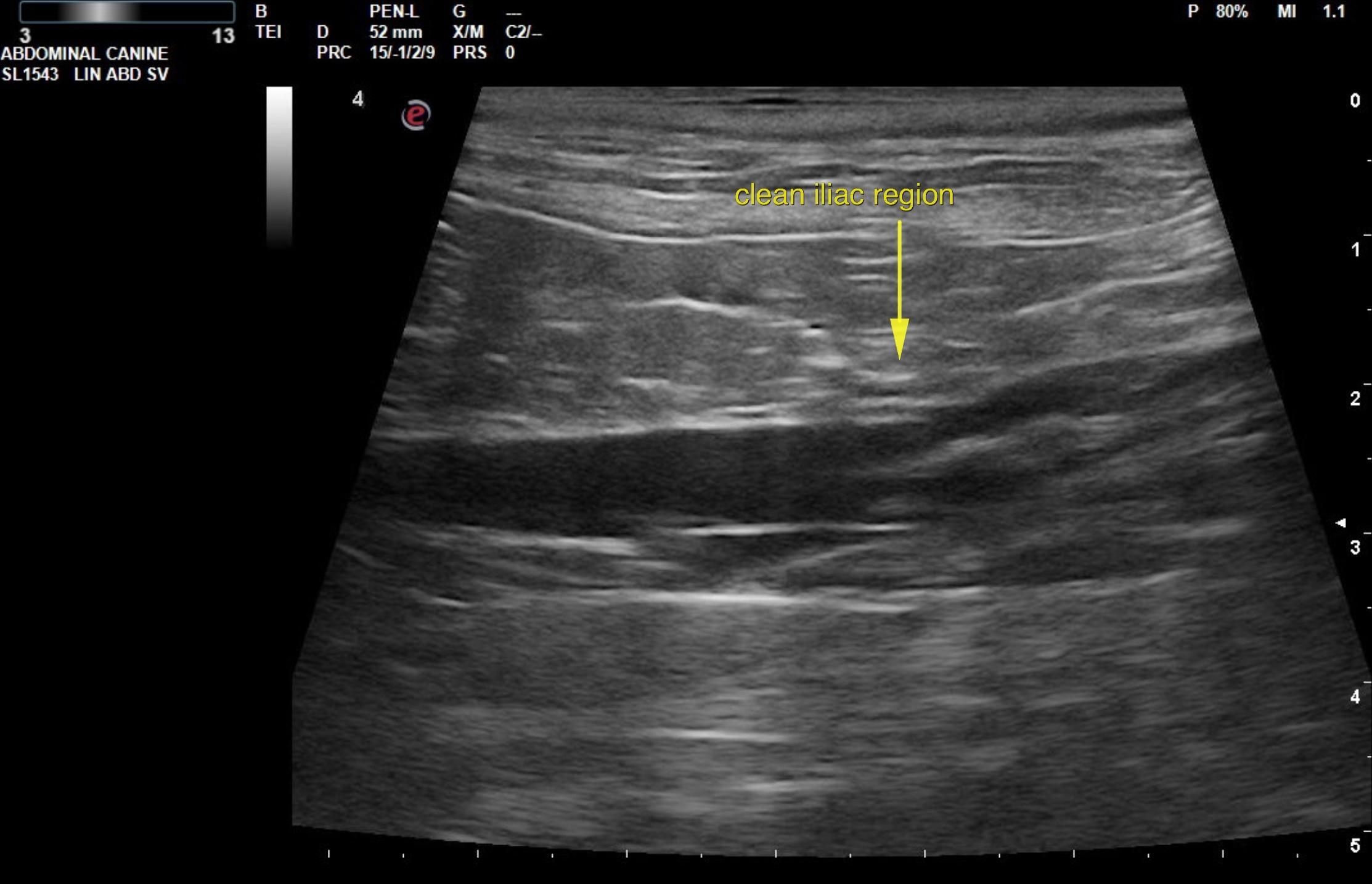
The iliac trifurcation was unremarkable.

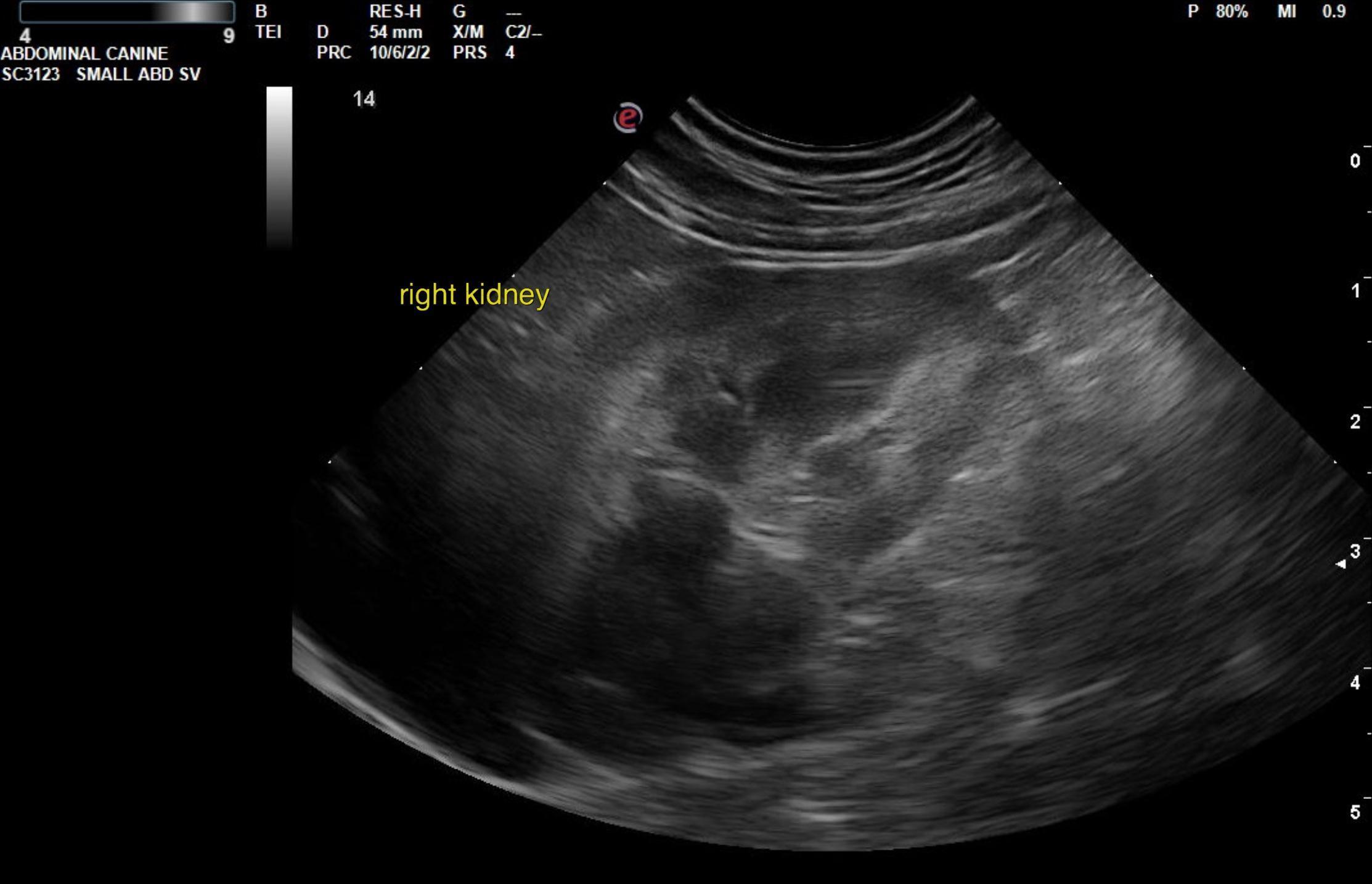
The kidneys revealed normal size and structure, corticomedullary definition and ratio for this age patient. The cortices presented largely uniform texture with normal echogenic relationship to liver and spleen. Medullary echogenicity differed distinctly from that of the cortex and no evidence or dilation could be seen. The capsules were acceptably uniform without dramatic irregularities. The right kidney measured 4.0 cm. 

DX

Chronic cystitis vs TCC vs round cell in the bladder

Outcome

Biopsies are strongly suggested in this patient, which can be cystoscopy guided to assess the urethra as well or if only surgical options are present, then biopsy of the ventral caudal aspect of the bladder wall, particularly that near the cystourethral junction would be recommended. These would ideally be guided by intraoperative ultrasound to assess the region of loss of detail. Chronic cystitis is still possible; however, transitional cell carcinoma and round cell neoplasia are also possible in this case. Culture and biopsy of the bladder is recommended. Guarded prognosis depending upon underlying histopathology. There was no evidence of perforation and no evidence of metastatic disease at this time.

Clinical Differential Diagnosis

Bladder – chronic cystitis, neoplasia, uroliths, polyploid cystitis
Urethra – neoplasia, lith, granulomatous urethritis

Patient Information

Patient Name : Lola Lein
Gender : Female, Spayed
Species : Canine
Type of Imaging : Ultrasound
Status : Complete
Liz Wuz Here : Yes
Code : 06_00359

Clinical Signs

  • Hematuria

Images

eli1eli2eli5eli6eli7

Blood Chemistry

  • Globulin, High

Clinical Signs

  • Hematuria

Urinalysi

  • Specific Gravity Low
Skip to content