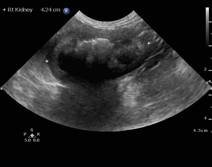
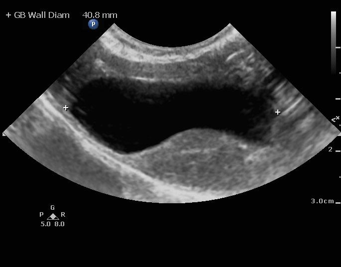
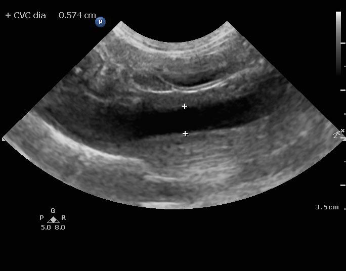
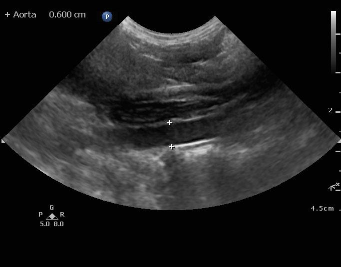
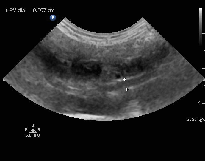
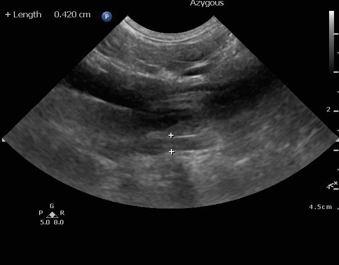
An 8-year-old spayed female Maltese dog was presented for evaluation of severely elevated pre-and post-prandial bile acids. In addition, the patient was positive for Rocky Mountain spotted fever. Seizure activity was present in the history
An 8-year-old spayed female Maltese dog was presented for evaluation of severely elevated pre-and post-prandial bile acids. In addition, the patient was positive for Rocky Mountain spotted fever. Seizure activity was present in the history