Splenoazygos Shunt in a 10 year old MN Schnauzer with bile acid elevation

Case Study

Splenoazygos Shunt in a 10 year old MN Schnauzer with bile acid elevation

This 10 year old MN Schnauzer dog has a history of low BUN from 10/2015. Elevated bile acids panel (pre 49, post 77). Rads show small liver, bladder stone. Asymptomatic.

This 10 year old MN Schnauzer dog has a history of low BUN from 10/2015. Elevated bile acids panel (pre 49, post 77). Rads show small liver, bladder stone. Asymptomatic.

Image Interpretation

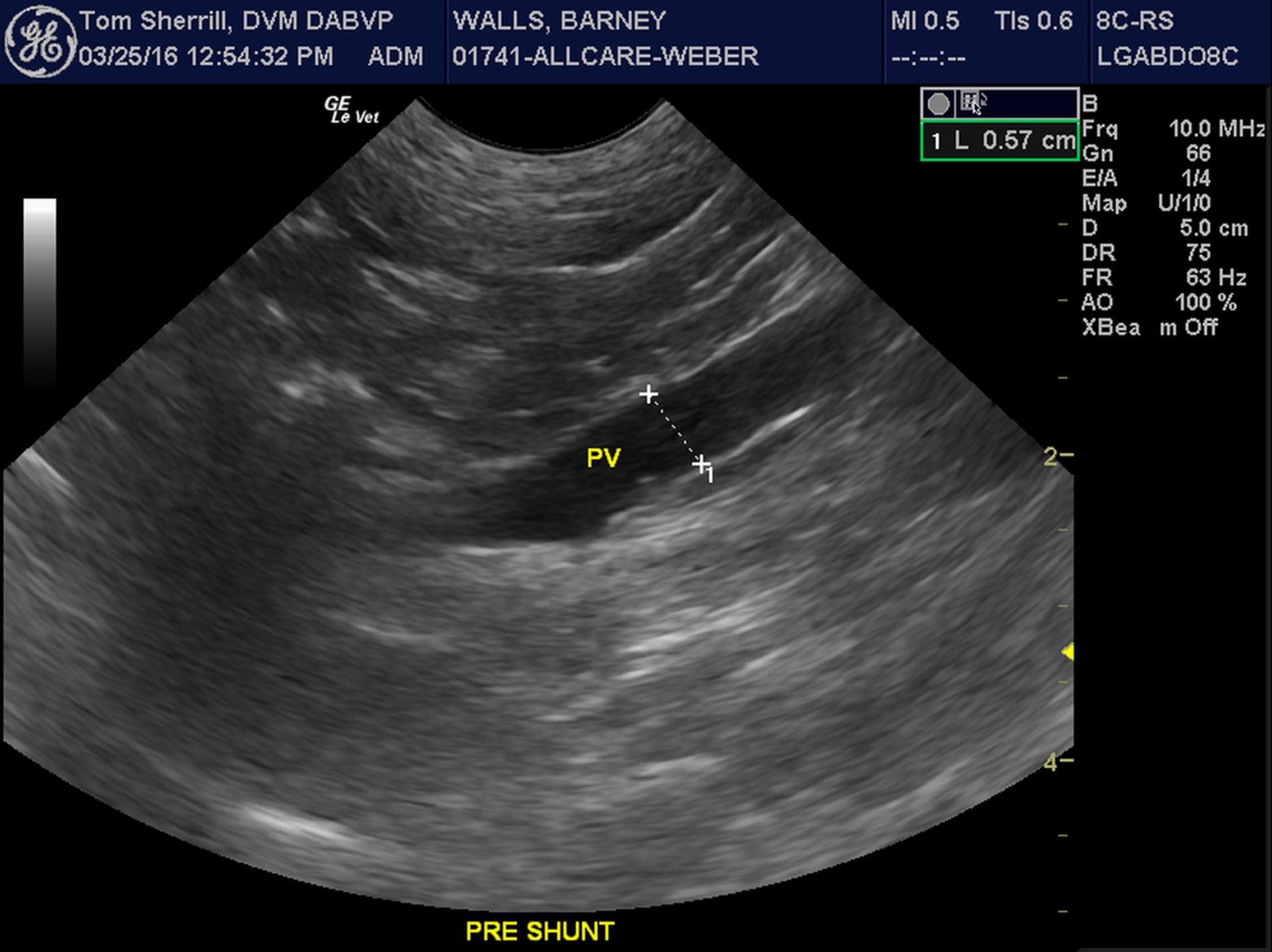
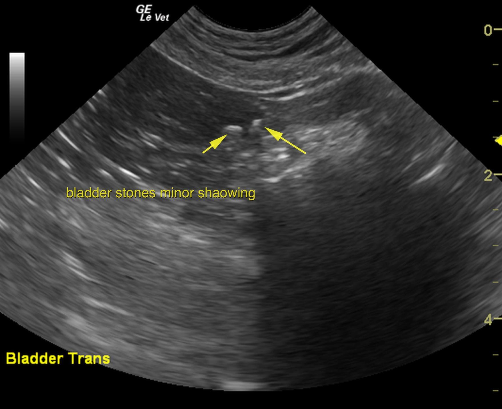
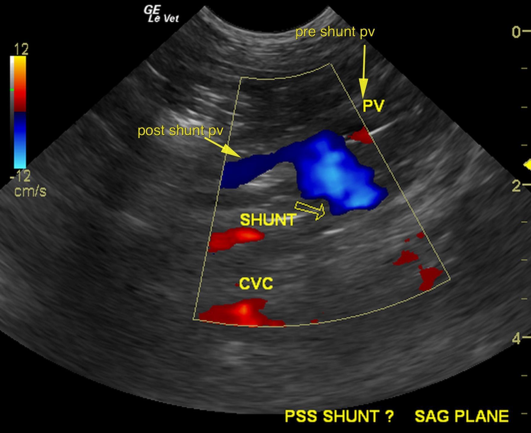
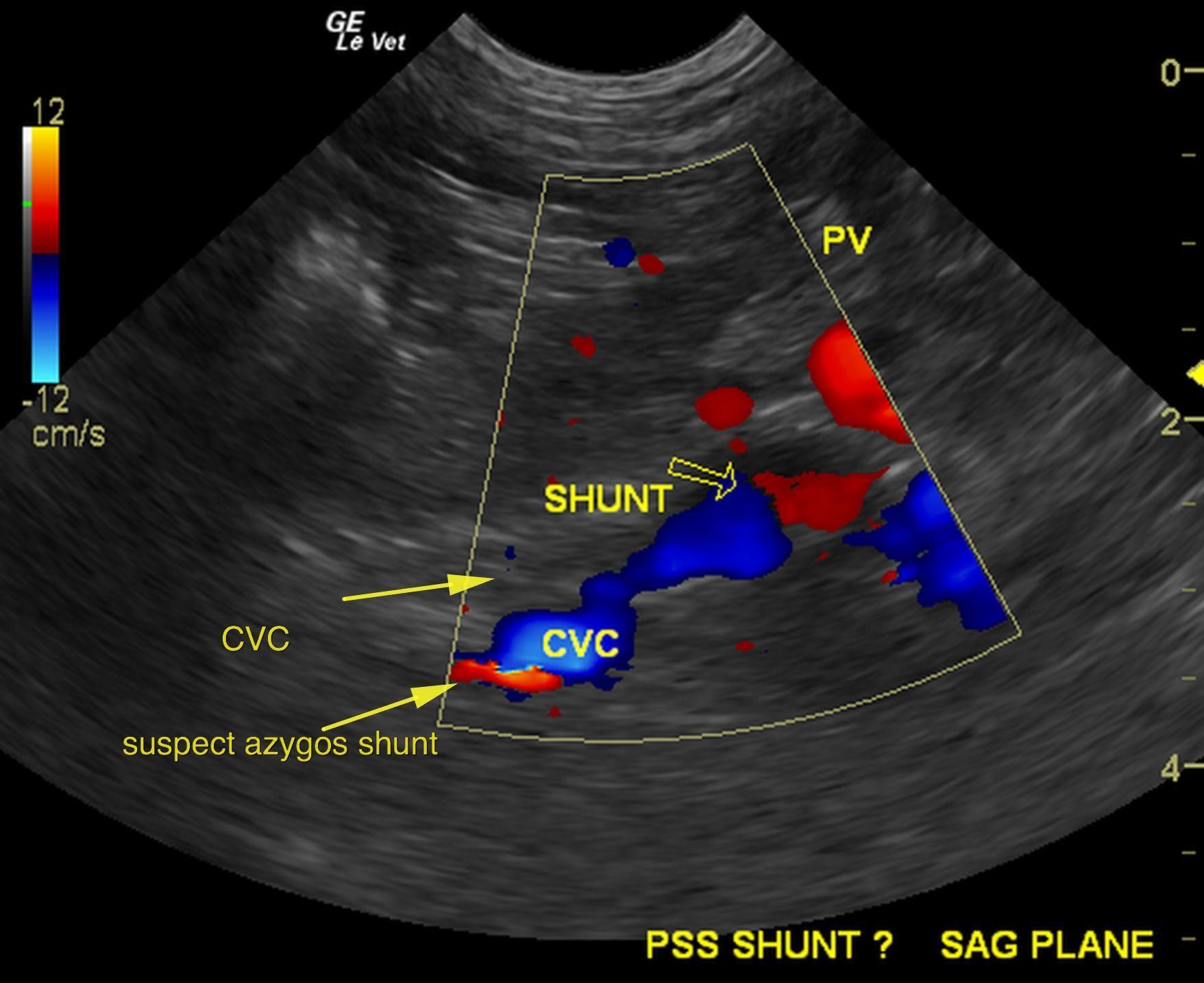
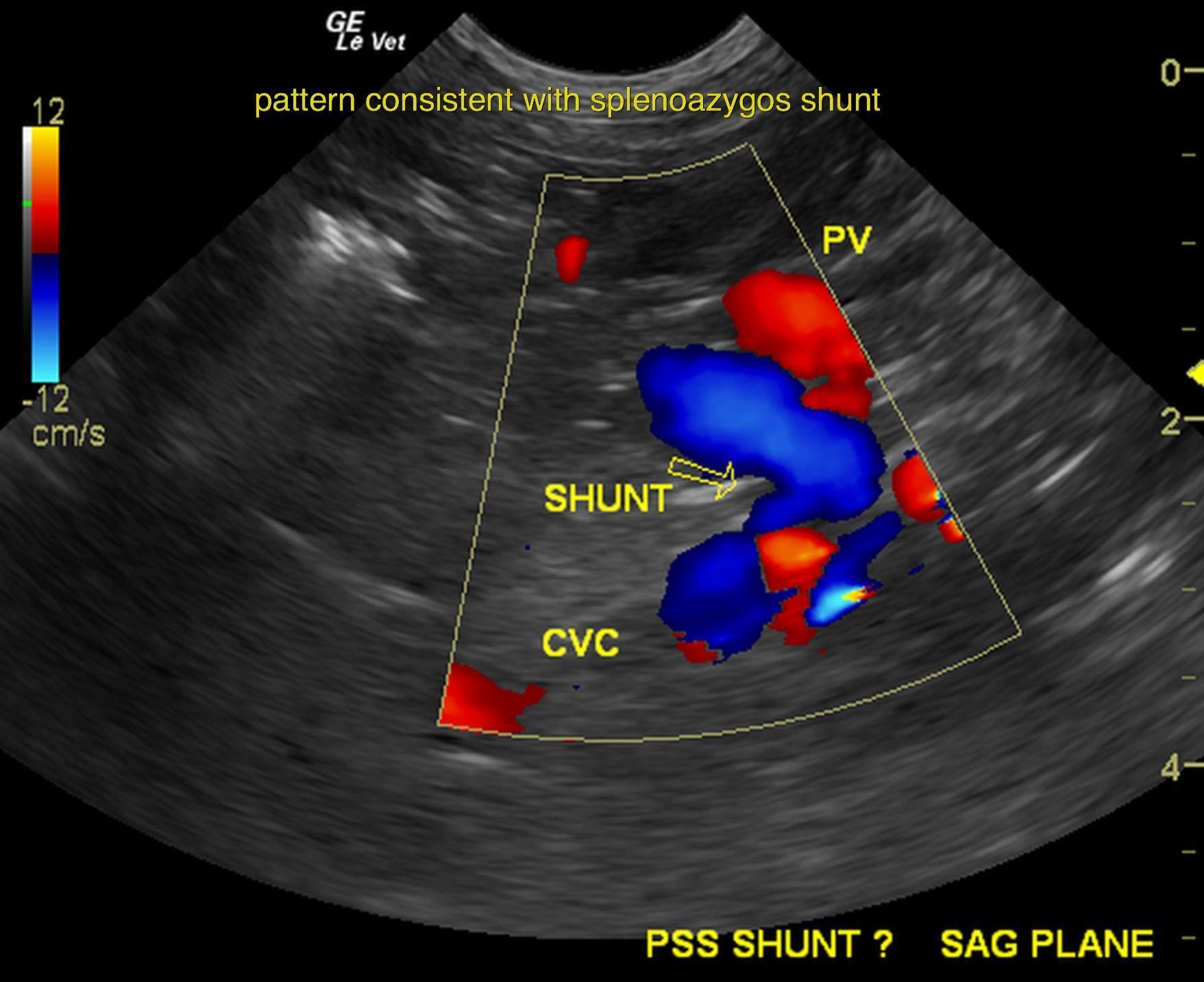
The bladder presented minor micropolypoid changes and small slight shadowing calculi. Bladder calculus measuring 0.2cm was also noted. Liver was subnormal in size with hypovascularity. Portal vein revealed a splenic vein derived shunt measuring approximately 0.8cm at the deviation from the portal vein. The portal vein post-shunt measured aporoximately 0.3cm; pre-shunt 0.57cm. The shunt appeared to discourse dorsally approximately 0.8-1.0cm in maximum width. The vena cava at the level of the diaphragm measured approximately 0.6cm. The contour of the shunt was most consistent with a splenoazygos shunt which would fit best with the elevated bile acids of 77. The contour of the shunt appears to be deriving from the splenic junction of the portal vein discoursing dorssally, the maximum of 0.8cm bypassng the vena cava and entering into the diaphragm dorsal tot he vena cava and ventral to the aorta, which could be the countour and pattern most consistent with splenozygos shunts.

DX

Splenoazygos shunt

Outcome

The presence of bladder calculi, microhepatica and the pattern of the blue and discoursing vessel would be most consistent with splenoazygos shunt. This ideally would be confirmed by CT evaluation with contrast or direct exploratory surgery with expectations towards ameroid constrictor ligation This does not appear to be a secondary shunt, but a primary shunt and splenoazygos shunts tend to be late manifesting and often have no significant liver values other than bile acid elevation.
The small calculi in the bladder would be suggestive for ammonium biurate. They do not appear obstructive at this time. It is debatable on whether cystotomy would be necessary. No evidence of secondary shunting or chronic liver disease was noted other than pure microhepatica with relatively unremarkable parenchyma.

Patient Information

Patient Name : Barney Walls/All Care Veterinary Hospital
Gender : Male, Neutered
Species : Canine
Type of Imaging : Ultrasound
Status : Complete
Liz Wuz Here : Yes
Code : 03_00555

Exam Finding

  • Microhepatica

Images

bar3bar4bar5bar7bar8

Blood Chemistry

  • BUN low
  • Post-Prandial Bile Acids, High
Skip to content