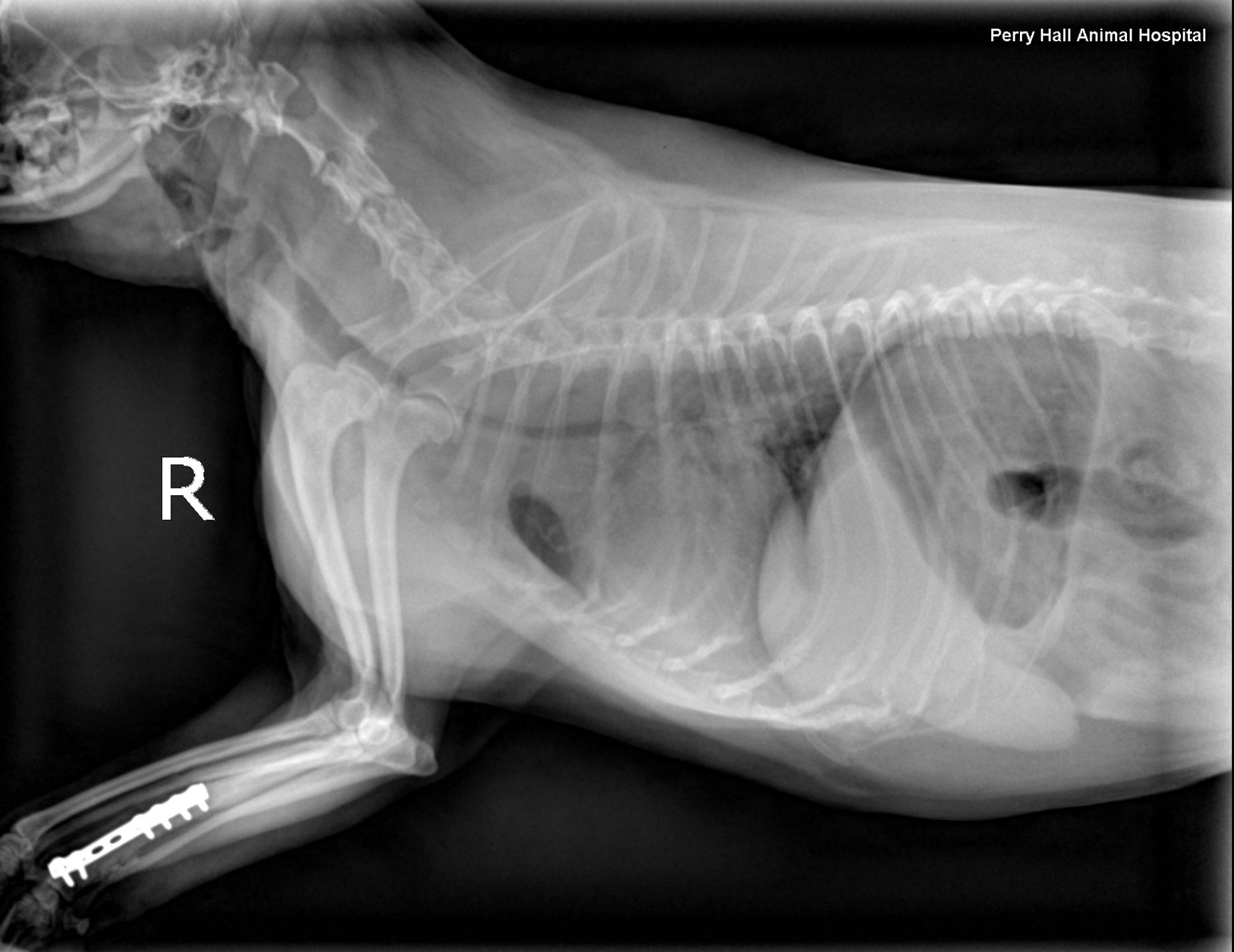
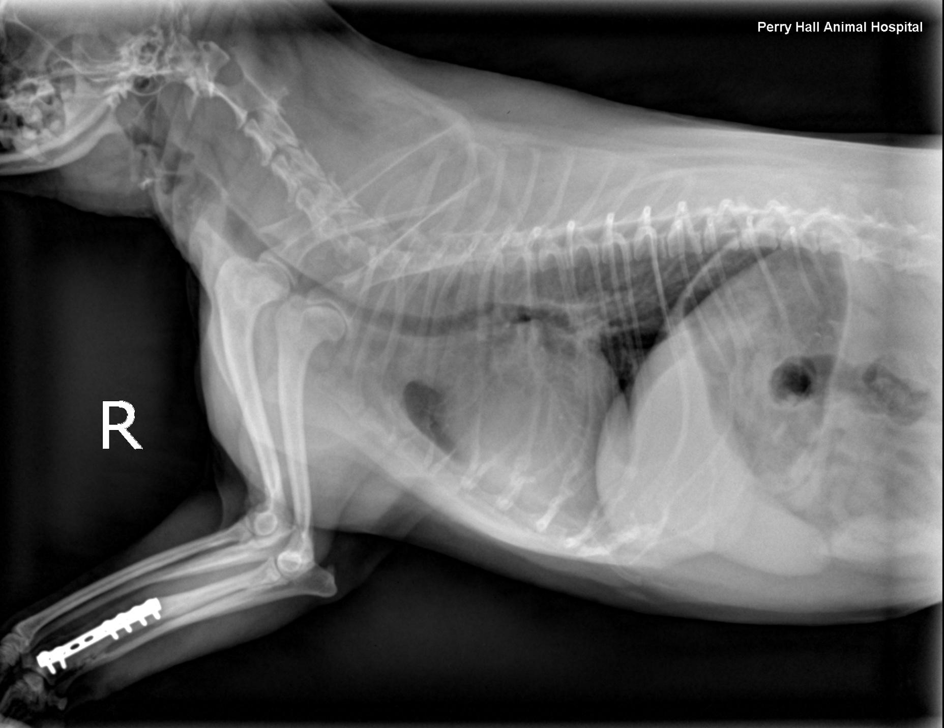
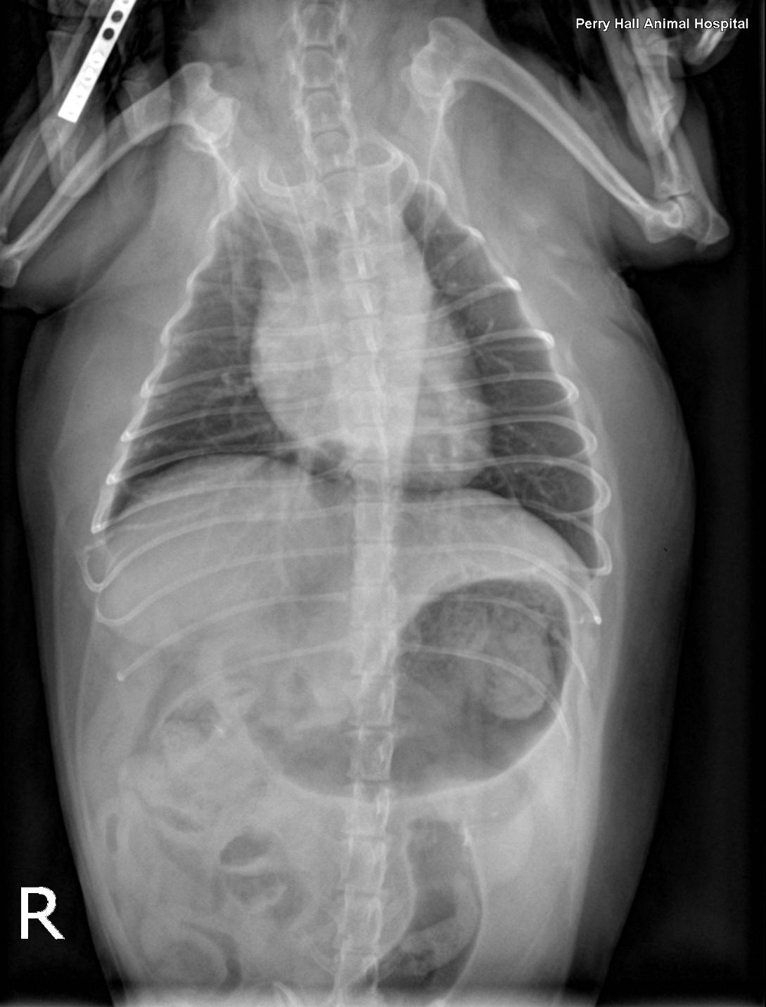
Rads- Left lateral, right lateral and VD views of the thorax and neck. One of the lateral views was obtained in fair inspiration (degree of inspiration reduced as a function of the disease), one was an expiratory view. There was mild to moderate rotation of the trunk in the lateral views. There was superimposition of the front limbs with the cranial thorax. The hands of the holding person were in the primary beam during exposure which is strictly prohibited.
The patient was obese.
Osseous structures: The patient showed a status after rightsided forearm fracture fixed with a DC plate in remodeling phase with radioulnar synostosis. There was mild bilateral shoulder osteoarthritis.
Extrathoracic soft tissue structures: The stomach was moderately distended with gas and food. The liver extended beyond the costal arch and mild lobar rounding was noted. There was a multilobulated soft tissue swelling with mass effect caudoventral to the mandibles.
Intrathoracic structures: The chest volume was small. There was an intrathoracic tracheal collapse and collapse of the main stem bronchi on the expiratory view. The height of the trachea was reduced by more than 50 % as compared with the inspiratory view. The tracheal lumen was reduced to a height of 2mm level with the thoracic inlet when collapsed. The cardiac silhouette was normal for size and shape. The major vessels were within normal limits. The pulmonary vessels and caudal vena cava were within normal limits. No mediastinal widening was noted.
The lungs showed diffuse increase in opacity caudodorsally on the expiratory view