RADS- Soft Tissue Mass Within the Region of the Aortic Bulge and Bronchointerstitial Pattern in an 11 year old MN Miniature Schnauzer dog

Case Study

RADS- Soft Tissue Mass Within the Region of the Aortic Bulge and Bronchointerstitial Pattern in an 11 year old MN Miniature Schnauzer dog

History of “abnormal breathing”. The patient was started on enalapril and lasix after radiographs on 5/18/15. Recheck radiographs on 7/15/15 revealed concern for a mass.

History of “abnormal breathing”. The patient was started on enalapril and lasix after radiographs on 5/18/15. Recheck radiographs on 7/15/15 revealed concern for a mass.

Image Interpretation

Rads of left lateral, right lateral and VD thorax – Osseous structures: within normal limits.
Extrathoracic soft tissue structures: There were multifocal small linear mineral opacities associated with the left renal diverticuli. There was no aerophagia.
Intrathoracic structures: The chest volume was normal. The course of the trachea was within normal limits. The overall size of the cardiac silhouette was within normal limits. There was an ovoid soft tissue opacity noted level with the aortic root which corresponded to mild focal mediastinal widening level with the aortic arch on the ventrodorsal view. No tracheal displacement was seen. The remainder of major vessels were within normal limits. The pulmonary vessels and caudal vena cava were within normal limits. The lungs showed a mild generalized bronchointerstitial pattern. There was tracheal and bronchial wall minerlization.

DX

soft tissue opaque mass effect within the region of the aortic bulge

Outcome

In general the following differential diagnoses have to be considered:
1. Age related normalcy
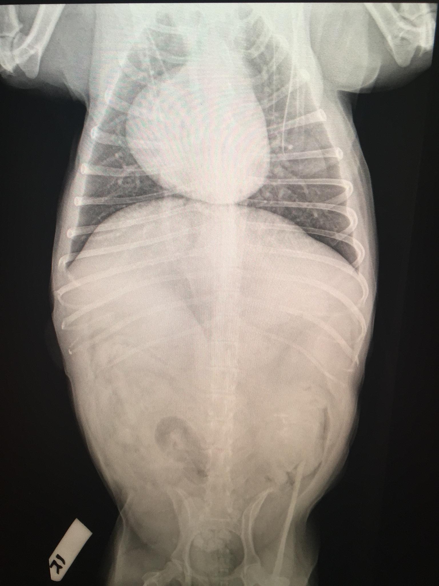
2. Poststenotic dilation or dilation due to systemic hypertension – the presence of subaortic/aortic stenosis has been ruled out by cardiac echo. The presence of systemic hypertension should be verified.
3. Aortic root aneurysm: spontaneous – unlikely because very rare, inherited collagenase defect such as Marfan-like syndrome – unlikely due to the age of the patient.
4. Aortic root paraganglioma, ectopic thyroid/parathyroid carcinoma at cardiac base, cranial mediastinal lymph node enlargement – theoretically an emerging neoplasia cannot be ruled out based on radiographs, but overall this is unlikely because no mass effect on trachea or specific mediastinal widening was present. Also an aortic root mass was not reported from the cardiac echo.
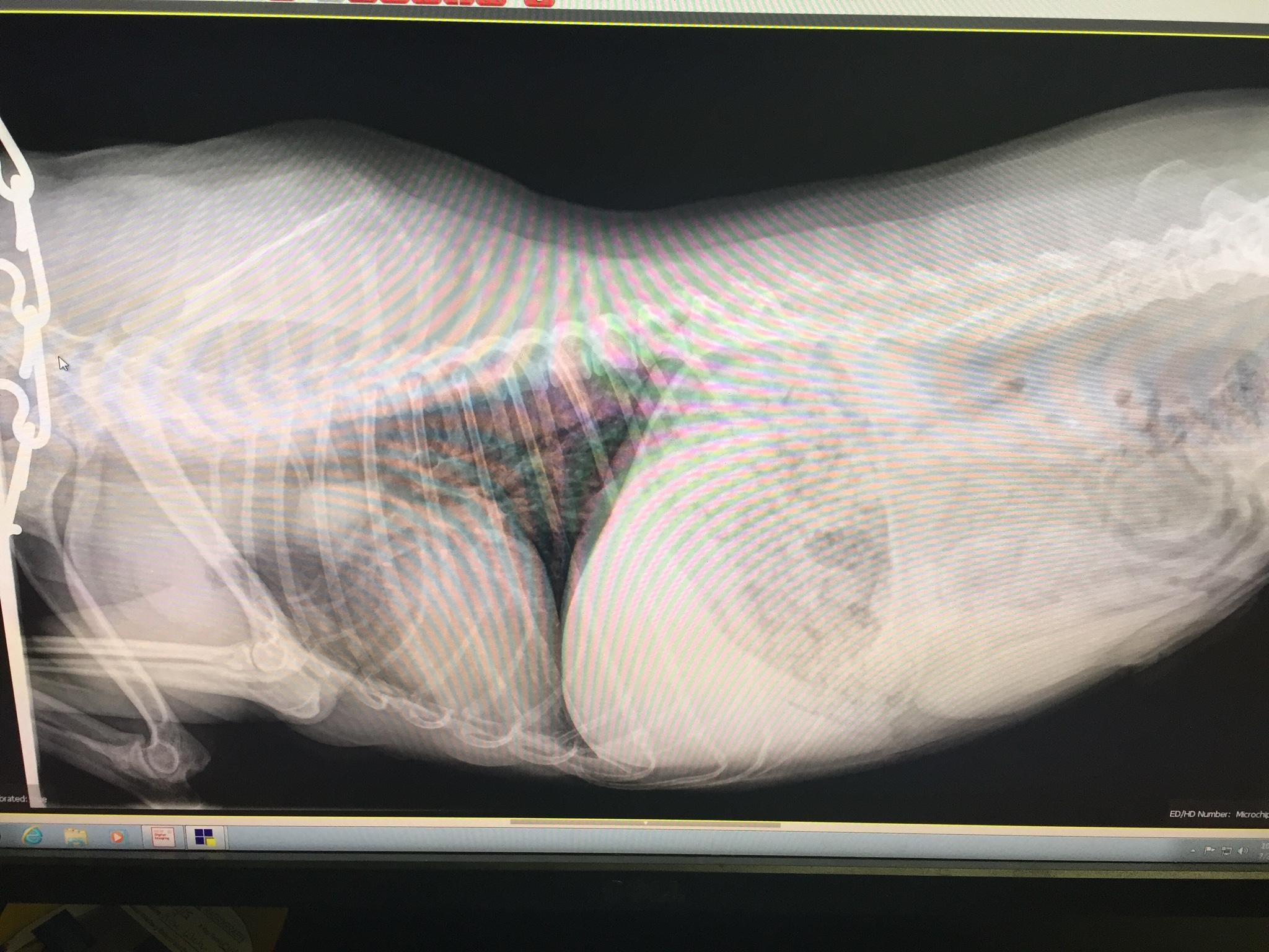
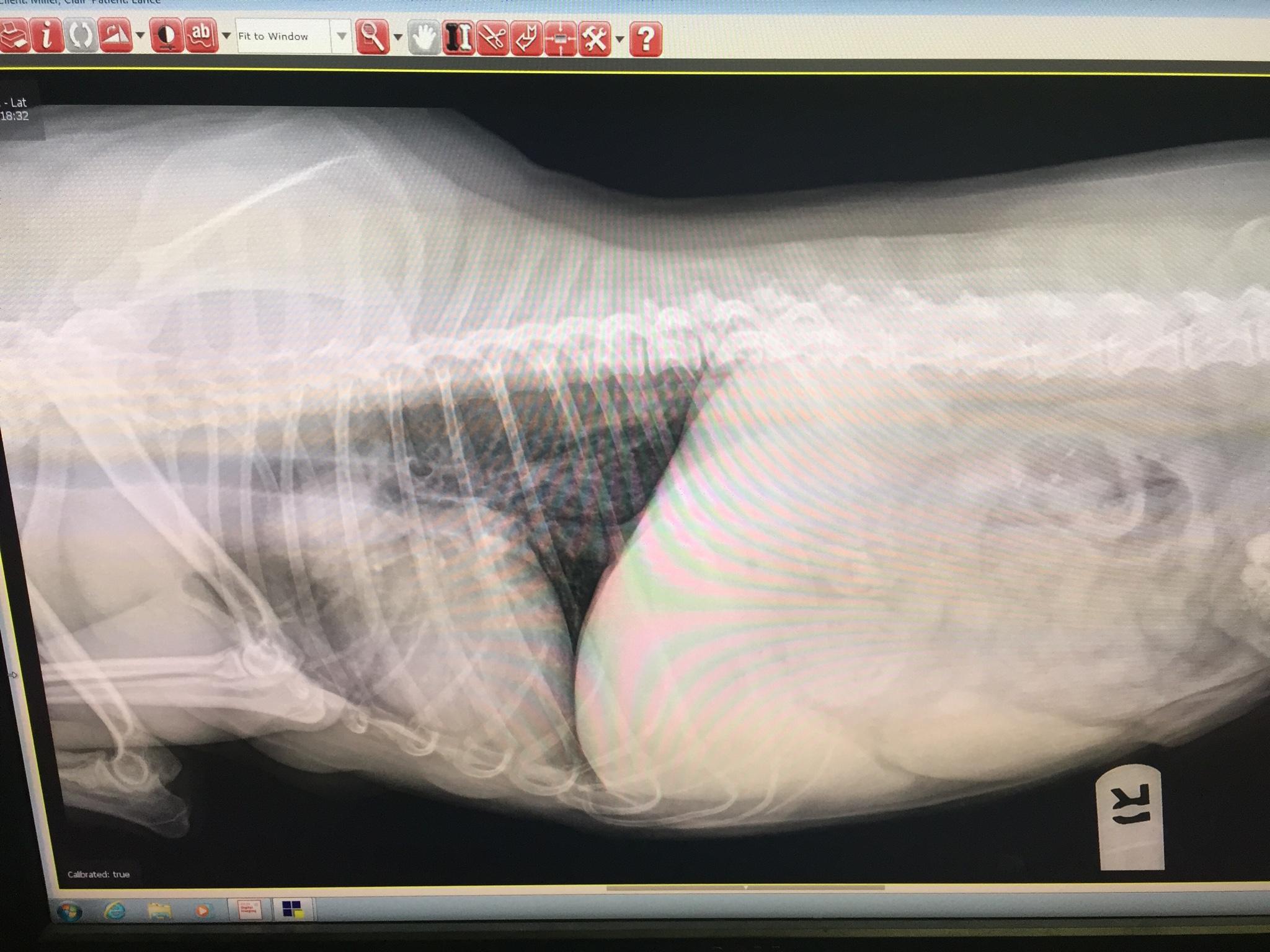
There was a mild bronchointerstital pattern which was likely age related. The presence of chronic infectious (bacterial/viral >> parasitic/fungal) or allergic bronchitis cannot be ruled out.
There was mineralization associated with the left renal diverticuli compatible with precipitation of crystals and/or renal stone formation within the renal pelvis.
The clinical relevance of the radiographic findings is questionable. Thoracic CT with contrast would be necessary to further evaluate the findings. Systemic disease associated with abnormal breathing has to be considered too.

Comments

 A total number of 3 views were available for assessment photographed from a screen ion jpeg format. There were artifacts from photographing the screen throughout the images. The degree of inspiration was fair. There was mild rotation of the trunk in all views. There was superimposition of the front limbs with the cranial thorax.


Patient Information

Patient Name : Lance Miller, Lehigh Valley
Gender : Male, Neutered
Species : Canine
Type of Imaging : Ultrasound
Status : Complete

Exam Finding

  • Respiratory Distress

Images

img_3824img_3825img_3826
Skip to content