RADS – Severe Tracheal Collapse in a 10 year old dog

Case Study

RADS – Severe Tracheal Collapse in a 10 year old dog

History of chronic cough, moist, seems tracheal.

Physical Exam: temp 102, inspiratory noises, HR-80. U/S-heart looks normal, la/ao-ok 

CBC and Chemistry WNL

History of chronic cough, moist, seems tracheal.

Physical Exam: temp 102, inspiratory noises, HR-80. U/S-heart looks normal, la/ao-ok 

CBC and Chemistry WNL

Image Interpretation

Right and left lateral and VD thorax: The patient was obese.
Osseous structures:
There were moderate degenerative changes associated with the axial skeleton. Both elbow joints presented mild osteoarthrosis.
Extrathoracic soft tissues:
The serosal detail was normal. The liver was mildly enlarged but normal for shape and opacity. There was moderate aerophagia.
Intrathoracic structures:
There was an ovoid gas opacity with even soft tissue opaque lining ventral to the
cranial thoracic trachea which was consistent with an esophageal redundancy.
There was a large amount of mediastinal fat.
The trachea presented severe focal narrowing spanning a distance of at least 4 cm level with the caudal neck and thoracic inlet. This was seen on both lateral views. The maximum degree of narrowing was noted level with the first intercostal space. The tracheal height was reduced by more than 90 % at that level. The tracheal narrowing was extended further by redundancy and infolding of the dorsal tracheal ligament cranial to the collapse.
The cardiac silhouette was within normal limits. The major and pulmonary vessels were within normal limits. The lung parenchyma presented a mild generalized bronchointerstitial pattern.

DX

The radiographic findings are compatible with a severe focal tracheal collapse level with the caudal neck and thoracic inlet. This is further accompanied by redundancy of the tracheal membrane.

Outcome

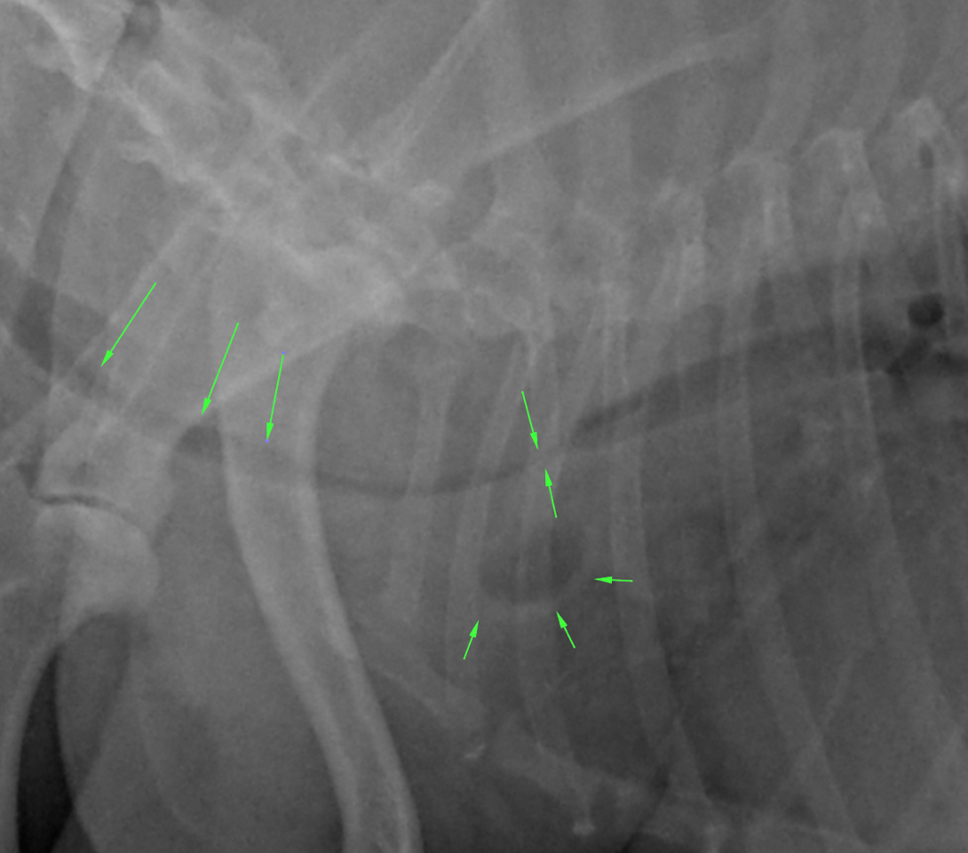
Note that generalized bronchomalacia with bronchial collapse is common in patients with tracheal collapse and cannot be ruled out radiographically.

A redundant cranial thoracic esophagus was noted which is a frequent finding in brachycephalic dogs. The overlap with the radiographic appearance of an esophageal diverticulum is referenced here but is very low for potential.

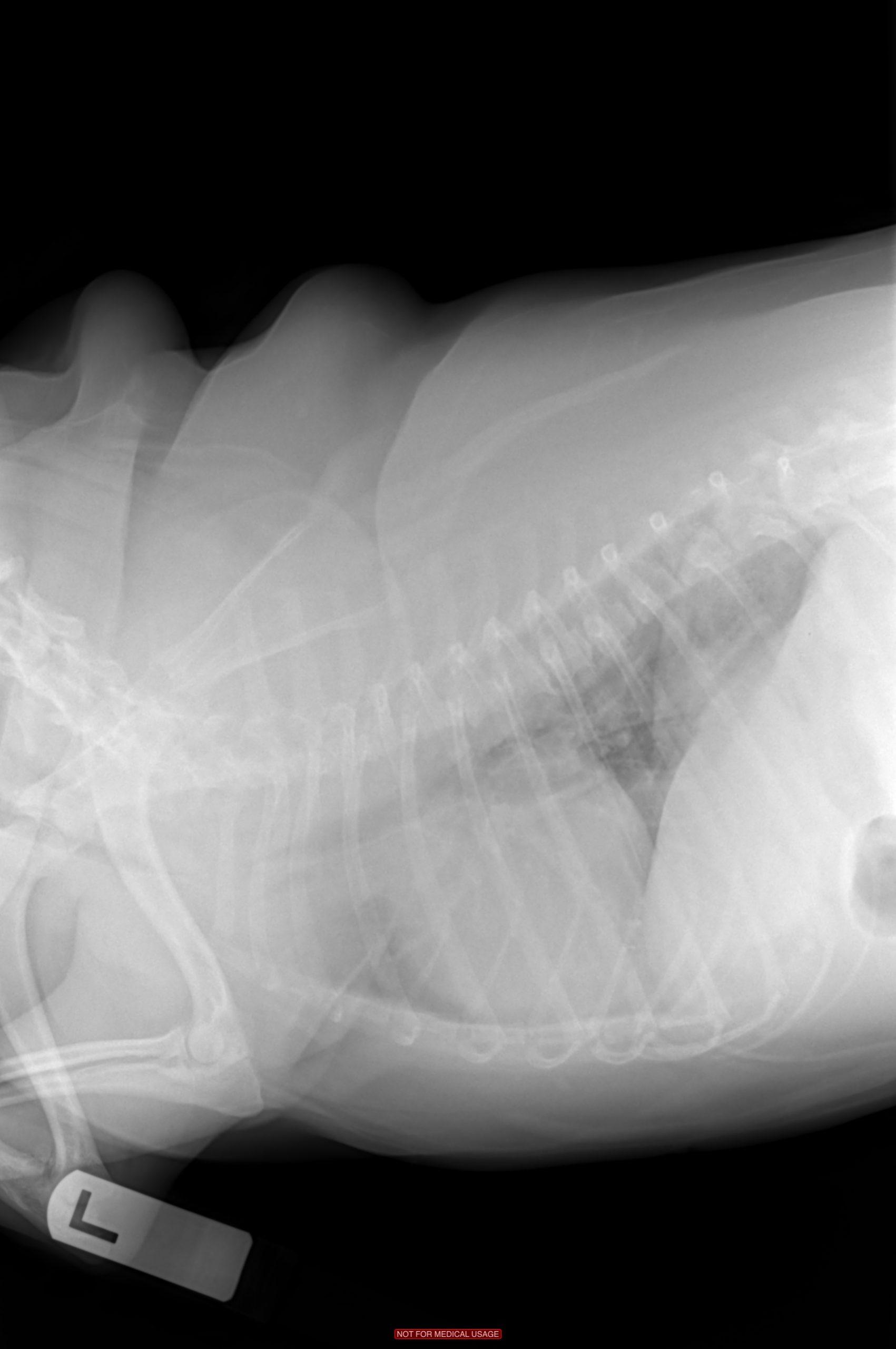
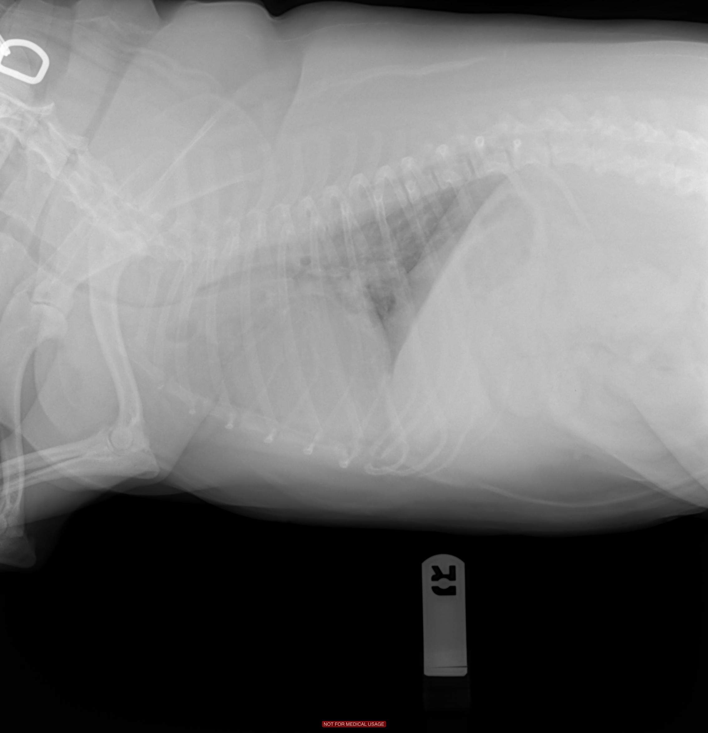
The aerophagia likely is a function of respiratory distress here.

The mild hepatomegaly is a frequent finding in patients with chronic respiratory distress and hypoxia.

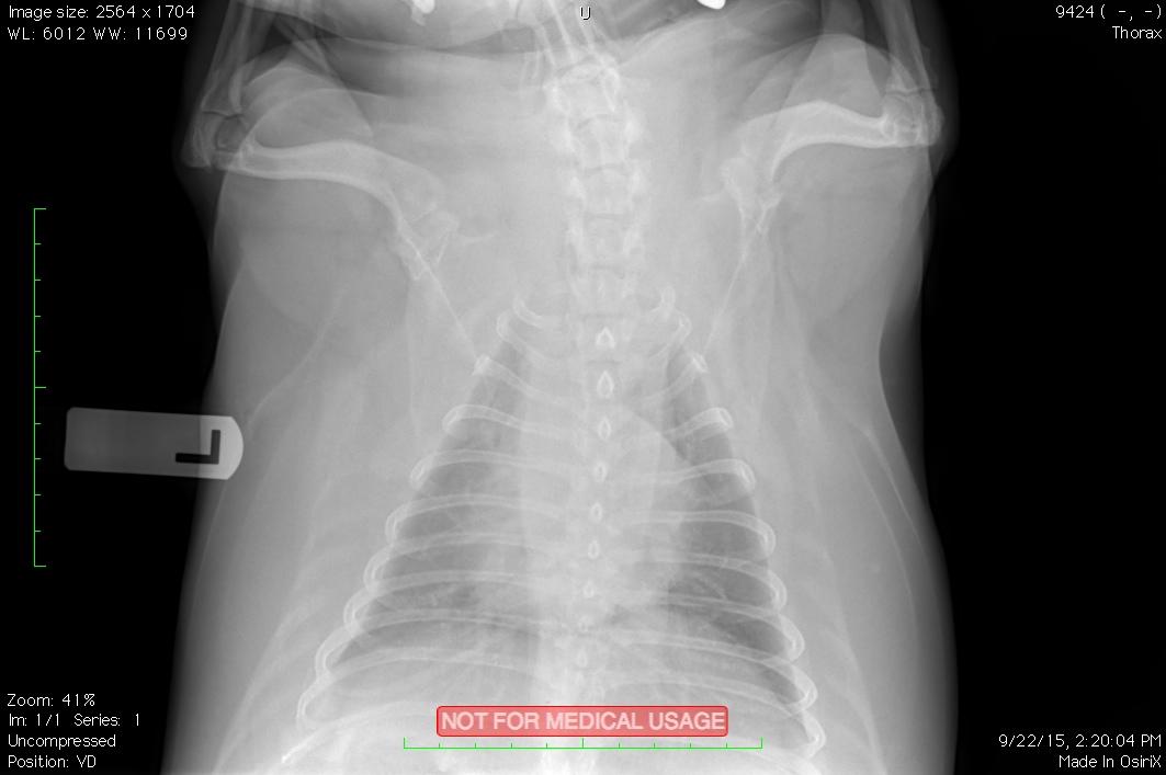
A mild bronchointerstitial lung pattern was noted which likely was an age related finding with no current significance.

Patient Information

Patient Name : Twista Rogers, Bayshore
Species : Canine
Type of Imaging : Ultrasound
Status : Complete

Clinical Signs

  • Coughing

Images

bildschirmfoto_2015-09-24_um_18twistaim-0001-0002im-0001-0003

Clinical Signs

  • Coughing
Skip to content