RADS – Chronic Upper Airway Obstruction with Chronic Inflammatory Airway Disease in a 9 year old MN Labrador Retriever dog

Case Study

RADS – Chronic Upper Airway Obstruction with Chronic Inflammatory Airway Disease in a 9 year old MN Labrador Retriever dog

Presented with roaring respirations. Physical exam: clinical signs suggestive of laryngeal paralysis. Inspiratory noise. CBC and chemistry WNL.

Presented with roaring respirations. Physical exam: clinical signs suggestive of laryngeal paralysis. Inspiratory noise. CBC and chemistry WNL.

Image Interpretation

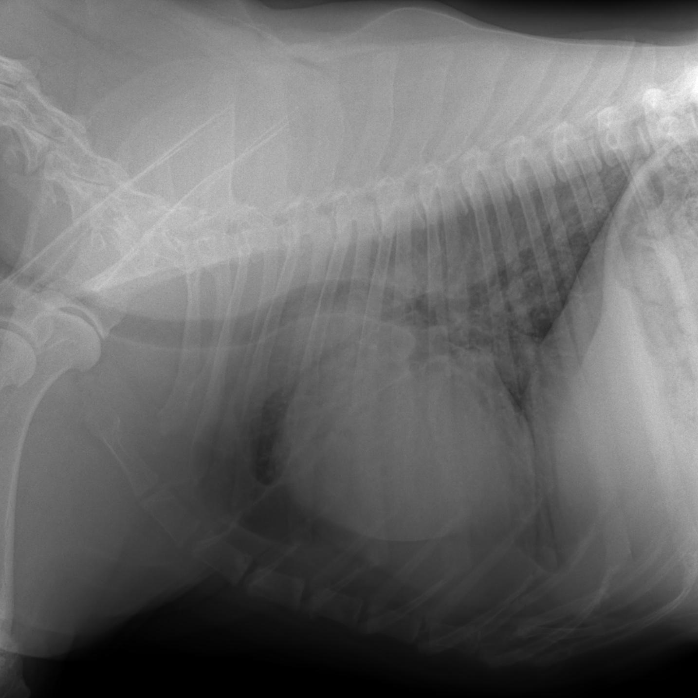
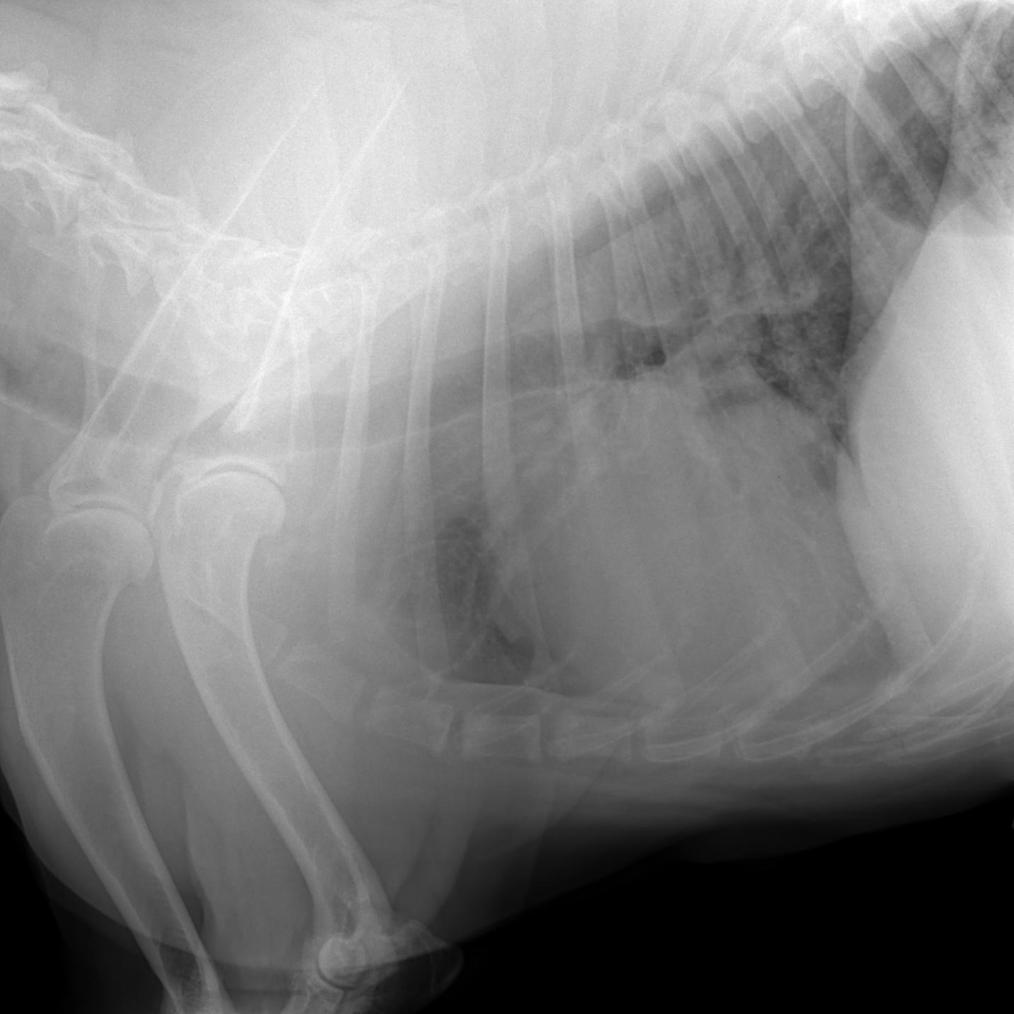
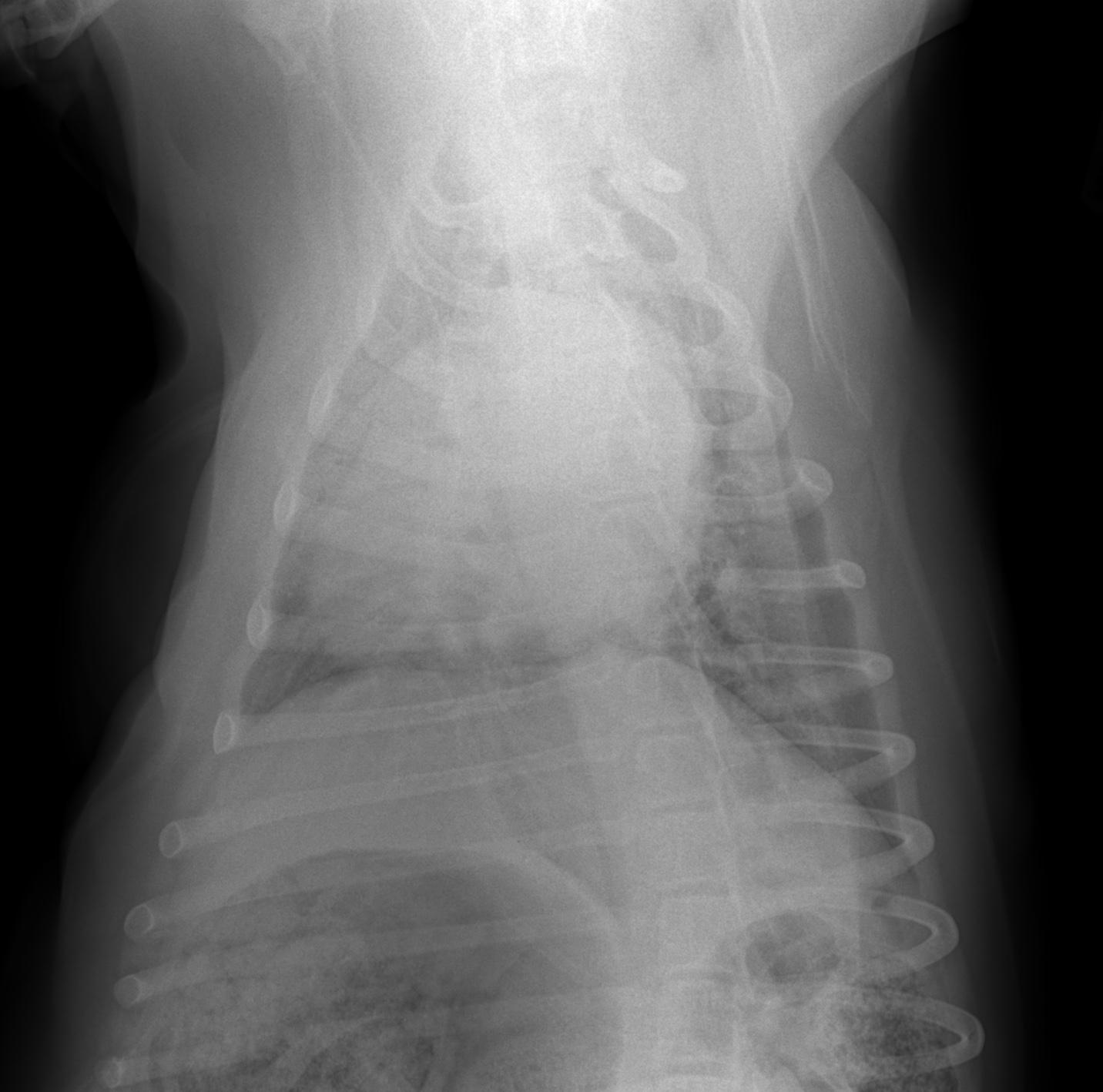
Rads – left lateral, right lateral and VD thorax: The degree of inspiration was fair to moderate. There wa moderate rotation on the VD view.
Osseous structures: the elbow joints showed moderate osteoarthrosis, there was minor osteoarthrosis of the shoulder joints. There was mild osteoarthrosis of the synovial vertebral joints throughout the cervical spine as imaged and mild spondylosis within the mid thoracic spine.
Extrathoracic soft tissue structures: There was a lipoma on the left chest wall. The stomach was moderately distended with gas and food. There was a redundant cervical tracheal membrane which usually is an incidental finding.
Intrathoracic structures: The chest volume was small. Even with moderate chest expansion and inspiration the diaphragmatic cupola remained in a cranial position and revealed pronounced cranial convex excursion. The cranioventral abdominal wall was midly tucked up.
The course of the trachea was normal – it showed dorsal bending on one of the lateral views as a function of the head position. The cardiac silhouette was normal for size and shape. The major vessels and pulmonary vessels were within normal limits. No mediastinal widening was noted.
The lungs showed a moderate generalized increase in opacity with a linear interstitial pattern, bronchial wall mineralization and peribronchial cuffing.

DX

The radiographic findings are compatible with a chronic incomplete upper airway obstruction which matches the clinical diagnosis of a laryngeal paralysis. Moreover there were radiographic signs of a chronic inflammatory airway disease. With the presence of upper airway disintegrity infectious bronchitis – especially viral and/or bacterial - is more likely in this case as compared with an allergic airway disease. Parasitic, fungal and mycobacterial infections are low for potential with the current radiographic presentation.

Outcome

For further diagnostic work up a bronchoscopy with bronchoalveolar lavage and lung worm testing are recommended prior to a potential surgical supply of the laryngeal disorder. There was no sign of aspiration pneumonia at this point. Aerophagia was seen as a function of dyspnea and/or stress.
A redundant tracheal membrane was noted. There was a chest wall lipoma, osteoarthrosis of the elbow, shoulder and cervical synovial joints and mild mid thoracic spondylosis. All are unrelated to the current clinical question.

Patient Information

Patient Name : Tucker Behrandt
Gender : Male, Neutered
Species : Canine
Type of Imaging : Ultrasound
Status : Complete

Exam Finding

  • Respiratory Distress

Images

bildschirmfoto_2015-07-23_um_11tucker_chest_radtucker_chest_rad_2tucker_chest_vd
Skip to content