lateral and DV thorax, right lateral neck – The patient was thin.
Osseous structures:
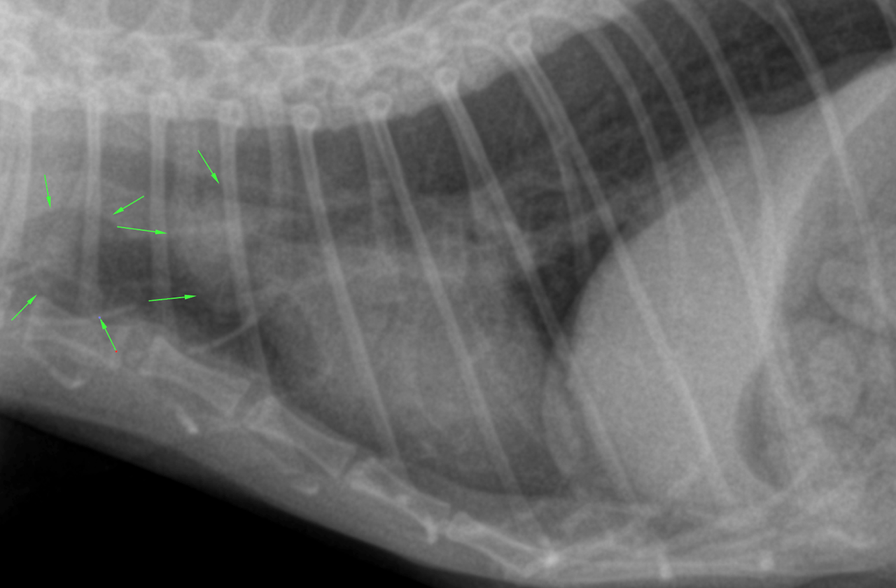
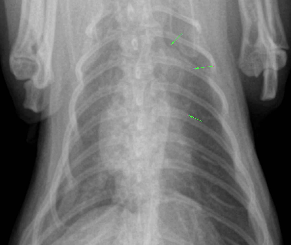
Mild degenerative changes were associated with the axial skeleton.
Extrathoracic soft tissues:
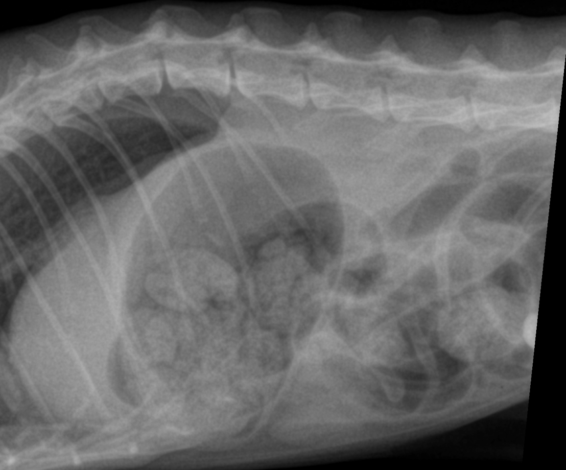
There was aerophagia with moderate amounts of gas within the stomach and intestine. The serosal detail was mildly reduced which likely was a function of lacking peritoneal fat.
Intrathoracic structures:
The esophagus was not seen. The course of the trachea was normal. The cardiac silhouette was within normal limits. There was a redundant aortic arch. The caudal vena cava and pulmonary vessels were thin. There was no mediastinal widening.
The chest was funnel shaped. The lung presented a mild generalized thinwalled bronchointerstitial pattern. There was no air trapping. There were no signs of expiratory obstruction. There were no signs of pneumonia, congestive heart failure, pulmonary edema or pleural effusion.