A 6-year-old MN Shih Tzu dog with history of cystotomy and increased liver enzymes, presented for Bile Acids panel. Markedly elevated pre prandial and markedly elevated post prandial results.
A 6-year-old MN Shih Tzu dog with history of cystotomy and increased liver enzymes, presented for Bile Acids panel. Markedly elevated pre prandial and markedly elevated post prandial results.
Case Study
Portosystemic shunt in a 6 year old MN Shih Tzu dog
Sonographic Differential Diagnosis
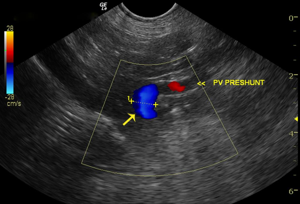
Extrahepatic portosystemic shunt consistent with splenoazygos (portoazygos) shunt.
Image Interpretation
The first image demonstrates a short dorsally directed vessel evidenced in color flow Doppler (Blue=away from transducer) deriving from the portal vein (splenic vein connection to the portal vein) indicating an extrahepatic shunt likely of splenic vein origin given the short length and position immediately dorsal to the portal vein. The dorsally directed contour would suggest termination either in the vena cava or the azygos vein. The second image of the portal hilus demonstrates the portal vein pre-shunt and post-shunt differential diameter; key to demonstrating the presence of a portosystemic shunt. Note the shunt diameter is 0.95 cm, pre-shunt portal vein 0.53 cm and post-shunt residual portal vein is 0.34 cm in diameter confirming the shunting of blood away from hepatic circulation. The short arrow demonstrates the well defined echogenic line of the diaphragm. The connection of the shunt (color flow Doppler blue) is beyond the diaphragm indicating an azygos vein connection (color flow Doppler red) indicated by the long arrow. Video 1 reveals a 1:1 ratio between the vena cava (near field) and aorta (far field) which is normal. hence the extrahepatic shunt must enter through the diaphragm and into the azygos vein in blue positioned in a vertical dorsally directed manner (Video 2, images 3, 4, and 5)
DX
Portosystemic shunt
Outcome
Lactulose therapy, metronidazole, and LD diet pending surgical repair of portosystemic shunt with biopsy of liver, was recommended for this patient. Patient underwent surgery to ligate portosystemic shunt and was later discharged with pain medication and L/D diet.
Clinical Differential Diagnosis
Liver pathology: portosystemic vascular anomaly (shunt, portal vein hypoplasia), microvascular dysplasia, hepatitis, hepatopathy, intestinal dysbiosis with falsely elevated bile acids
Sampling
Extrahepatic shunt confirmed at surgery.
Video
Patient Information
Patient Name :
Milo C
Gender :
Male, Neutered
Species :
Canine
Type of Imaging : Ultrasound
Status :
Complete
Liz Wuz Here :
Yes
Code :
03_00099
History
- Cystotomy
- Elevated Liver Enzymes
Images


Blood Chemistry
- ALT (SGPT), High
- AST (SGOT), High
- Post-Prandial Bile Acids, High
- Pre-Prandial Bile Acids, High