A 4-year-old F Yorkshire Terrier dog was presented at an emergency facility for acute onset disorientation, unresponsiveness, and vomiting. On physical examination, she appeared blind and was disoriented. Abnormalities on CBC and blood chemistry were low MCV and hypocholesterolemia. The patient was treated with K/D diet. Recheck blood work a month later showed alkaline phosphatase normalized and a normal WBC. Eight months later, the owner reported that the patient was still vomiting 1-2 times a week and getting Lactulose. CBC showed low MCV and MCHC.
A 4-year-old F Yorkshire Terrier dog was presented at an emergency facility for acute onset disorientation, unresponsiveness, and vomiting. On physical examination, she appeared blind and was disoriented. Abnormalities on CBC and blood chemistry were low MCV and hypocholesterolemia. The patient was treated with K/D diet. Recheck blood work a month later showed alkaline phosphatase normalized and a normal WBC. Eight months later, the owner reported that the patient was still vomiting 1-2 times a week and getting Lactulose. CBC showed low MCV and MCHC. The only change on blood chemistry was elevated ALP activity. Nutrical was added to the therapy. Two weeks later, the patient was presented for vomiting. On physical examination, the patient was febrile and slightly disoriented. The patient was treated with metronidazole and L/D diet. The following morning the owner reported that the patient was back to normal.
Case Study
Portoazygos shunt with renal and bladder calculi in a 4 year old F Yorkshire Terrier dog
Sonographic Differential Diagnosis
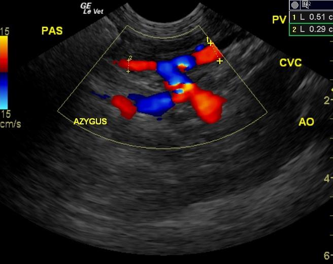
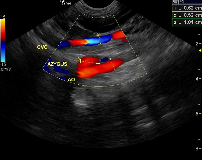
Portoazygos shunt. Significant renal calculi. Minor urinary bladder calculi.
Image Interpretation
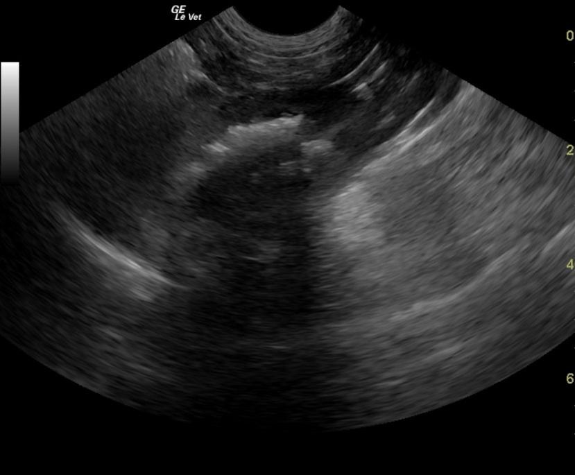
Small cystic calculi (at 0.4cm) were noted with shadowing in the urinary bladder (not shown). Image 1: The kidneys presented swollen contour with multiple corticomedullary calculi some of which were dramatic and obstructing the renal pelvis. The liver presented moderately subnormal size with 1.8cm of width in short axis. A 0.6cm portoazygos shunt was noted in this patient arising from the splenoportal region bypassing the vena cava and entering into the azygos vein at the level of the aorta prior to the diaphragm. The portal vein prior to the shunt measured 0.37cm after the shunt at 0.25cm, the vena cava measured 0.52cm in the post shunt region and the aorta measured 0.6cm in the post shunt region.
DX
Outcome
Surgical correction of a portocaval shunt was discussed. In the interim she was treated with lactulose, metronidazole, and L/D diet. Unfortunately, this patient did not make it to surgery; as the owner found her drowned in their neighbor’s pool a few weeks later.
Clinical Differential Diagnosis
Liver pathology: hepatic encephalopathy, portocaval shunt, chronic liver disease.
Video
Patient Information
Clinical Signs
- Disorientation
- Unresponsiveness
- Vomiting
Exam Finding
- Disorientation
Images
Blood Chemistry
- Cholesterol, Low
CBC
- MCV, High
Clinical Signs
- Disorientation
- Unresponsiveness
- Vomiting