A 10-year-old M Beagle was referred for ultrasonography after presenting for history of hypercalcemia, elevated cholesterol, and dysuria. A parathyroid panel was within normal limits.
A 10-year-old M Beagle was referred for ultrasonography after presenting for history of hypercalcemia, elevated cholesterol, and dysuria. A parathyroid panel was within normal limits.
Case Study
Parathyroid neuroendocrine neoplasm in a 10 year old MI Beagle
Sonographic Differential Diagnosis
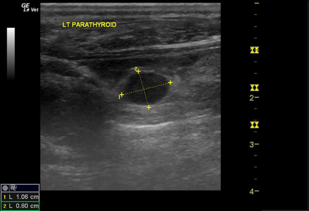
Left parathyroid adenoma.
Image Interpretation
The thyroid glands themselves were echogenic and micronodular. The possibility of hypothyroidism should be assessed. The left mid thyroid region revealed a 1.1cm hypoechoic nodule. This is in the region of the parathyroid and could represent an endocrine tumor. Fine needle aspirates were performed with 22 and 25 gauge needles in order to attempt to arrive at a cytological diagnosis.
DX
Neuroendocrine neoplasm, parathyroid
Outcome
A biopsy of the parathyroid gland to differentiate between an adenoma and adenocarcinoma was advised. No further outcome provided.
Clinical Differential Diagnosis
Humoral hypercalcemia of malignancy (HHM), hypoadrenocorticism, chronic kidney disease, hypervitaminosis D, and primary hyperparathyroidism. Less common etiologies include bone neoplasia, osteomyelitis, hypertrophic osteodystrophy, granulomatous disease, calcium supplementation, and oral phosphate binders.
Sampling
An US-guided FNA of the parathyroid gland revealed a neuroendocrine neoplasm.
Video
Patient Information
Patient Name :
Shiloh C
Gender :
Male, Intact
Species :
Canine
Type of Imaging : Ultrasound
Book :
yes
Status :
Complete
Liz Wuz Here :
Yes
Code :
13_00009
Clinical Signs
- Dysuria
Images
Blood Chemistry
- Calcium, High
- Cholesterol, High
Clinical Signs
- Dysuria