MRI – Meningocerebral herniation after craniotomy and chronic GME in a 10 year old MN Boston Terrier

Case Study

MRI – Meningocerebral herniation after craniotomy and chronic GME in a 10 year old MN Boston Terrier

This 10 year old MN Boston Terrier was previously diagnosed with granulomatous meningoencephalitis (GME). Presented with seizure activity.

This 10 year old MN Boston Terrier was previously diagnosed with granulomatous meningoencephalitis (GME). Presented with seizure activity.

Image Interpretation

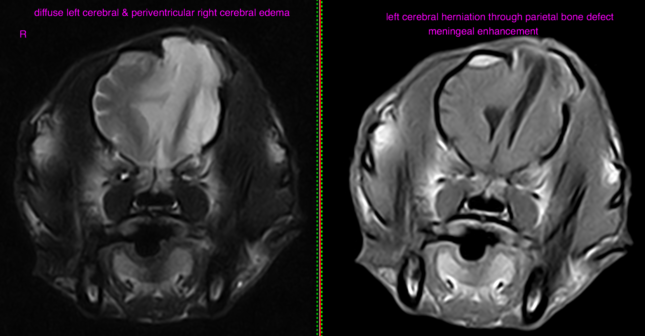
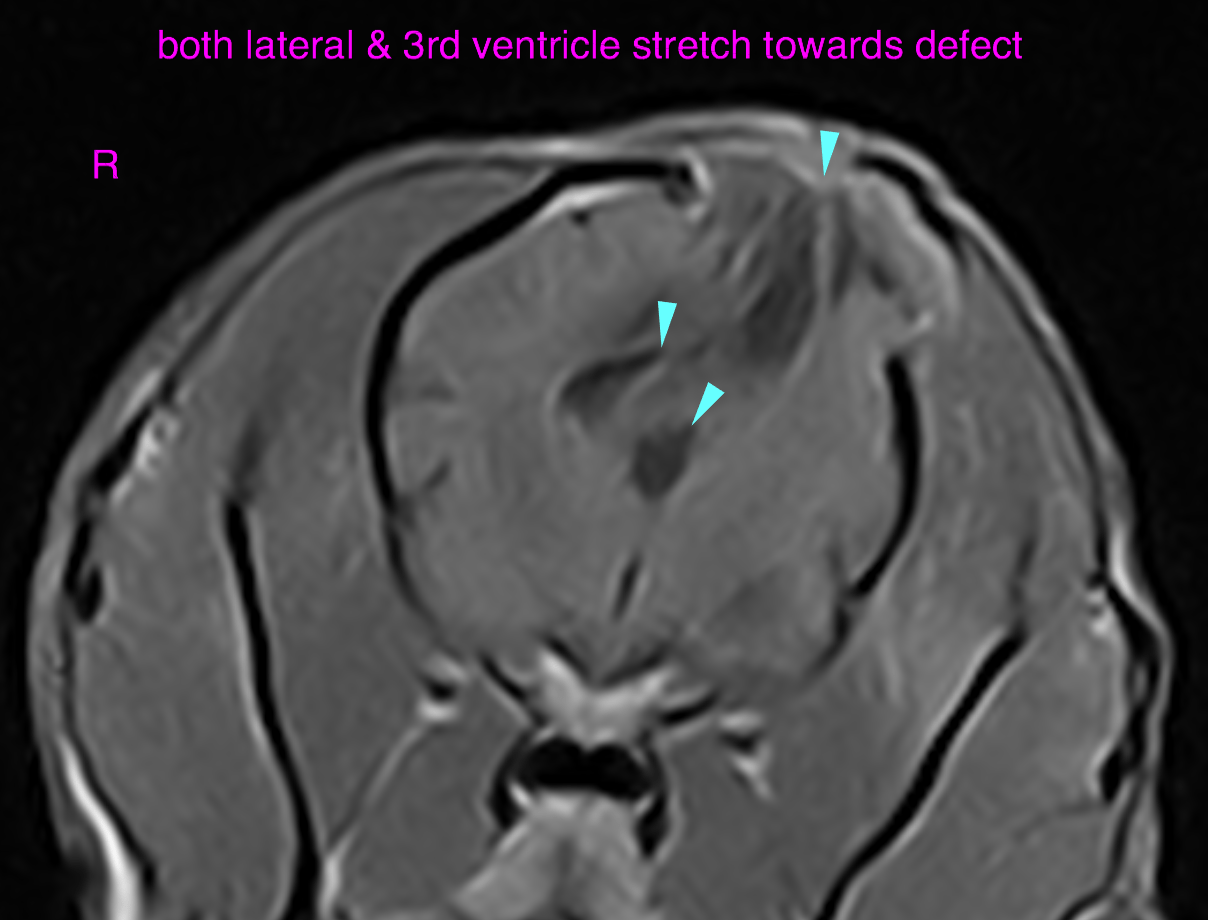
MRI – A 2 cm wide, well-delineated piece of bone is elevated dorsally from the left parietal bone (assumed iatrogenic from prior craniotomy). Part of the left cerebral hemisphere including the parietal lobe and the lateral ventricle are herniated through the bone defect. The brain parenchyma, both lateral ventricles and the third ventricle present longitudinal stretch towards the defect. The cerebral falx is shifted to the left. The entire left cerebral hemisphere presents diffuse T2-hyperintense edema. Extensive periventricular ill-defined white matter T2-hyperintensity is seen within the right cerebral hemisphere. 

DX

• Meningocerebral herniation after assumed craniotomy and chronic GME • mildly regressive brain edema • mild cerebral brain atrophy • meningeal and ependymal thickening compatible with meningitis versus scarring

Outcome

Both the meningoencephalitis and the surgical procedure have likely contributed to the meningitis.
There is no evidence of new granulomatous lesions.

Compared with the study dating 06/20/2016 the findings are slightly less pronounced.

Patient Information

Patient Name : Bacchus Barba/CVC
Gender : Male, Neutered
Species : Canine
Type of Imaging : Ultrasound
Status : Complete
Liz Wuz Here : Yes
Code : 20-00001

Clinical Signs

  • Seizures

Images

bildschirmfoto_2016-07-28_um_18bildschirmfoto_2016-07-28_um_18

Clinical Signs

  • Seizures
Skip to content