A 5-year-old MN poodle cross dog that was originally presented with an infection of unknown origin and hyperglobulinemia was now evaluated for anorexia and ADR. CBC showed leukocytosis and monocytosis. Urine specific gravity was normal (1.057).
A 5-year-old MN poodle cross dog that was originally presented with an infection of unknown origin and hyperglobulinemia was now evaluated for anorexia and ADR. CBC showed leukocytosis and monocytosis. Urine specific gravity was normal (1.057).
Case Study
Lymphoid Hyperplasia in a 5-year-old MN poodle cross dog
Sonographic Differential Diagnosis
Chronic inflammatory bowel disease with lymphadenitis. Chronic intestinal dysbiosis or parasitic disease as cause of underlying globulins. Mild potential for neoplasia.
Image Interpretation
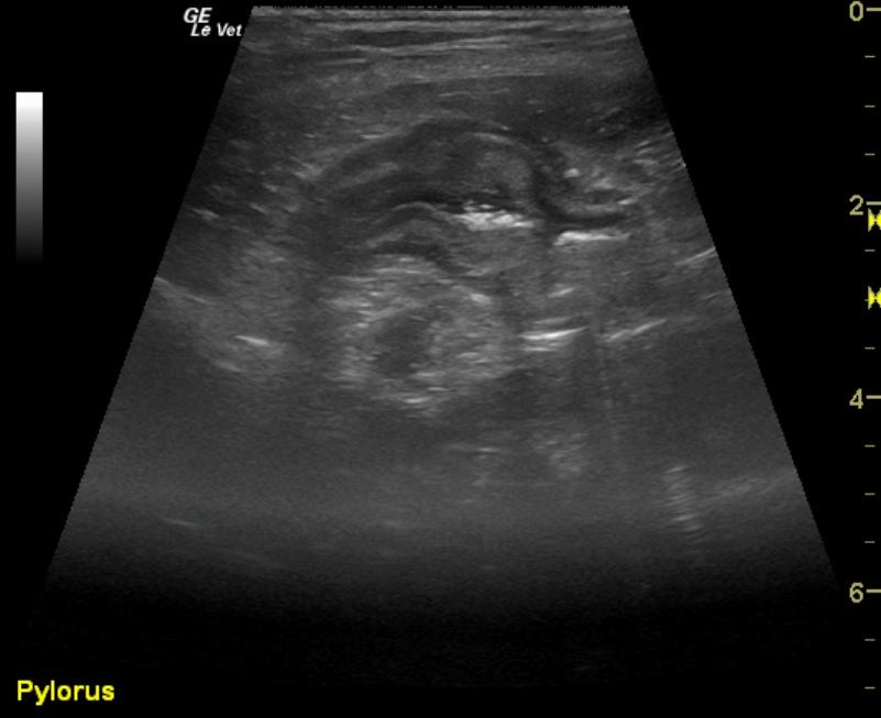
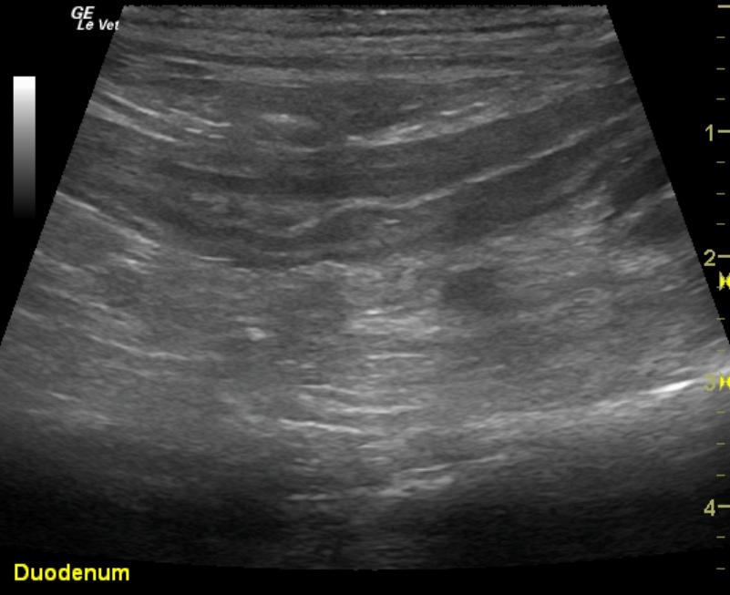
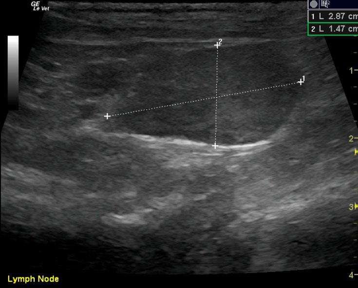
Pyloric outflow was mildly prominent in this patient with hypertrophy of the muscularis and mildly increased echogenicity of the submucosa layer. The remainder of the intestinal tract revealed thickened submucosal layers. Jejunal lymph nodes were enlarged at 2.87 x 1.47 cm with uniform contour, and the egg shape formation would be more suggestive of reactive lymphadenitis.
DX
Lymphoid hyperplasia
Outcome
The patient was still not 100% after 1 month of antibiotics, inflammatory leukogram 2% bands. Recheck ultrasound revealed the caudal abdominal lymph nodes were persistently enlarged, yet less reactive and measured 2.84 x 1.07 cm. They were uniformly, prominently echogenic. Iliac lymph nodes were also unremarkable at this time compared to the prior to sonogram and measured 1.22 x 0.47 cm on the right. Structurally this patient appears improved with minor residual mesenteric lymphadenopathy. Continuation of the current protocol would be recommended unless the patient demonstrates persistent clinical signs.
Comments
Antiparasitic protocol and dietary change along with Zithromax and metronidazole would be recommended over the next 2-3 weeks depending on clinical signs.
Clinical Differential Diagnosis
Multiple organ pathology -liver, spleen, pancreas, lymph nodes, kidney, peritoneal cavity (infection, inflammation, neoplasia) ; GI pathology (obstruction with focal peritonitis); Pyothorax
Sampling
FNAs were performed without evident complication, and showed lymphoid hyperplasia neutrophilic inflammation
UA Specific Gravity Range
1.057
Video
Patient Information
Patient Name :
Mardi H
Gender :
Male, Neutered
Species :
Canine
Type of Imaging : Ultrasound
Status :
Complete
Liz Wuz Here :
Yes
Code :
04_00292
Clinical Signs
- "Not Doing Right"
- Anorexia
Images
CBC
- Monocytes, High
- WBC, High
Clinical Signs
- "Not Doing Right"
- Anorexia