A 12-year-old MN Basset Hound was presented for evaluation of acute vomiting and painful abdomen.
A 12-year-old MN Basset Hound was presented for evaluation of acute vomiting and painful abdomen.
A 12-year-old MN Basset Hound was presented for evaluation of acute vomiting and painful abdomen.
A 12-year-old MN Basset Hound was presented for evaluation of acute vomiting and painful abdomen.
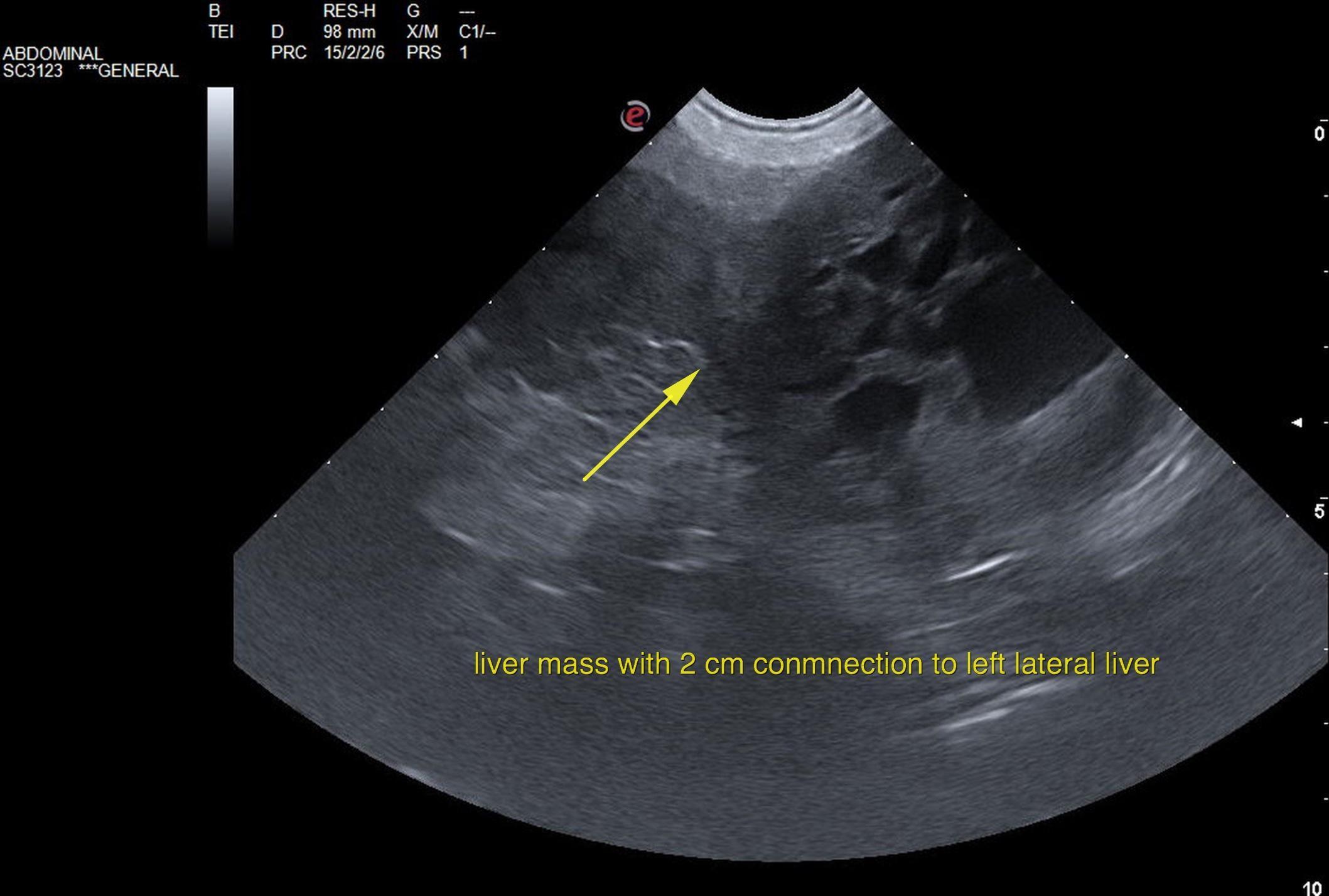
Left lateral liver mass; appears resectable. No overt evidence of metastatic disease.
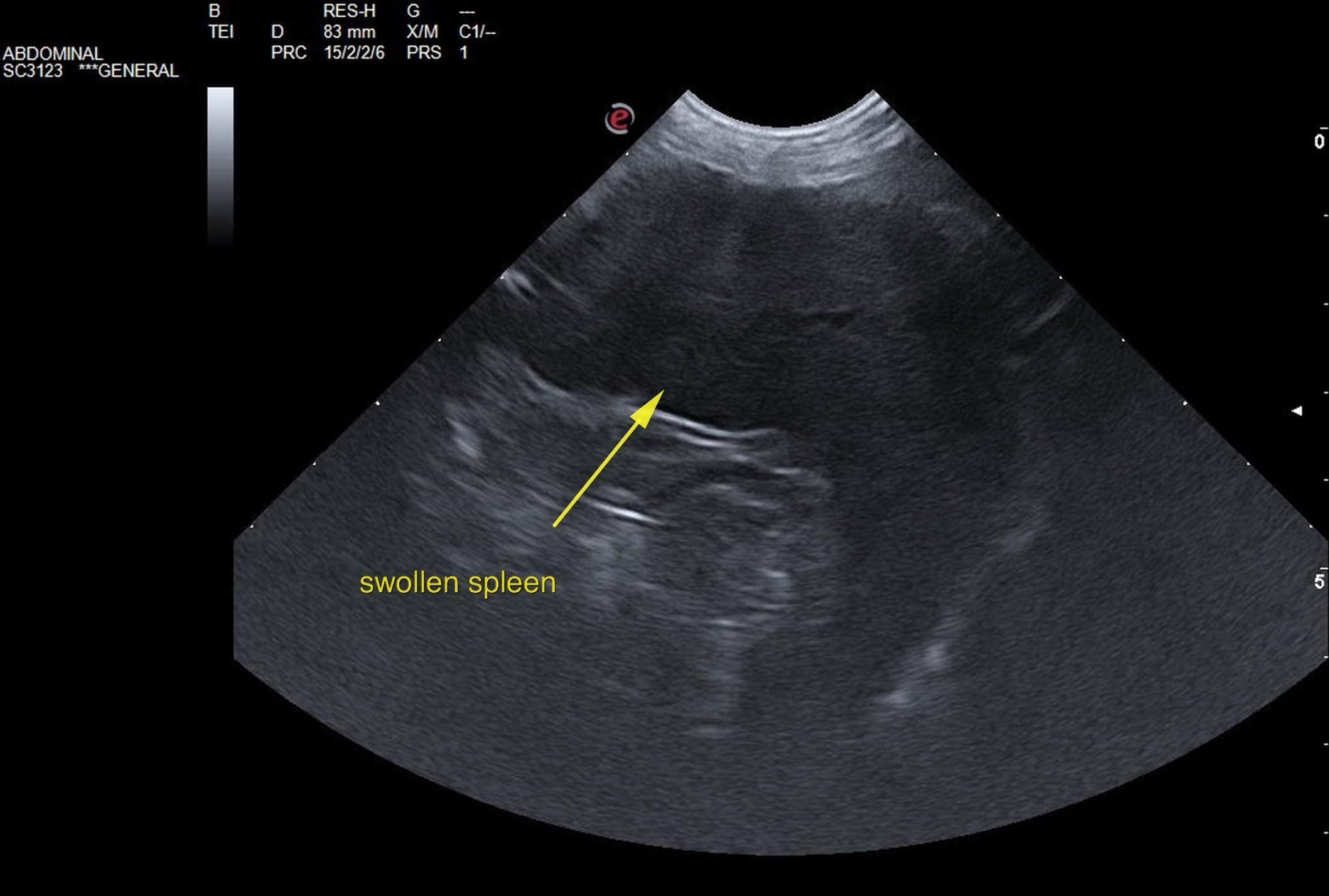
Hypersplenism splenic pattern. Inspection at surgery and potential splenectomy may be optimal in this patient.
The left lateral lobe of the liver revealed a mass connected to the caudal aspect of the left lateral liver lobe by a bridge of approximately 2 cm. This is at high potential for torsion given the pedunculated structure. Mild inflammatory pattern was noted around the liver mass; could not rule out lobe torsion given the inflammation. This is a surgical urgency. The remainder of the liver was unremarkable. Gallbladder presented minor polypoid changes. No evidence of other neoplasia.
The spleen was uniformly enlarged with relatively uniform parenchyma without evidence of masses. The capsule was mildly swollen. This is most consistent with hypersplenism and reactive hyperplasia deriving from splenic white or red pulp. However, early infiltrative disease, such as lymphoma or mast cell neoplasia can, at times, present in this manner.
US-guided FNA would be best in order to ensure only reactive hyperplasia is present. If clinical signs fit with potential neoplasia or mast cell disease, then Benadryl injection (1 mg/pound IM) 15 minutes prior to FNA would be recommended.
3-view chest radiographs are recommended. Left lobectomy is recommended with inspection of the spleen and judgment call at the time of surgery for splenectomy. I am not suspecting a neoplastic process in the spleen; however, it is enlarged and mildly pathological.
Peritonitis
Liver – acute hepatitis, neoplasia, lobe torsion
Gall bladder – cholecystitis
Pancreas – pancreatitis, neoplasia
Spleen – splenitis, torsion